Figure 71.2 M-mode recording of the mitral valve illustrating the high-frequency fluttering (arrows) of the anterior leaflet characteristic of aortic regurgitation due to aortic valvular flail. BP, blood pressure.
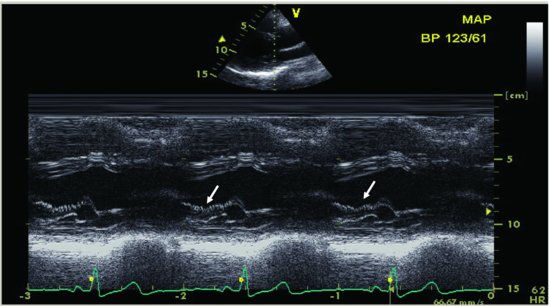
Figure 71.3 Suprasternal notch long-axis view illustrates the reversal flow in the aortic arch (a); pulse Doppler recording shows the forward and reversal flow in the abdominal aorta, which indicates severe aortic regurgitation due to aortic valvular flail (b). AAO, ascending aorta; ABDA, abdomen aorta; DAO, descending aorta.
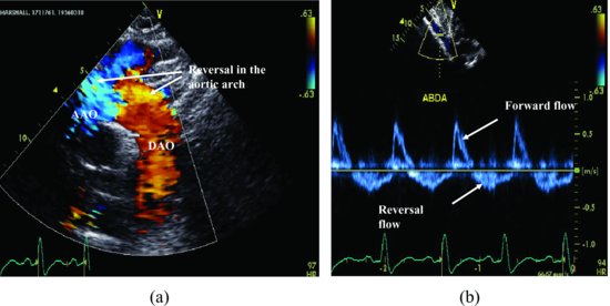
Figure 71.4 M-mode recording of parasternal long-axis view with color Doppler demonstrates the wide opening of aortic closing line and aortic regurgitant flow persistent in the entire diastolic period, which indicates severe aortic regurgitation.
Stay updated, free articles. Join our Telegram channel

Full access? Get Clinical Tree


