, Marilyn J. Siegel2, Tomasz Miszalski-Jamka3, 4 and Robert Pelberg1
(1)
The Christ Hospital Heart and Vascular Center of Greater Cincinnati, The Lindner Center for Research and Education, Cincinnati, OH, USA
(2)
Mallinckrodt Institute of Radiology, Washington University School of Medicine, St. Louis, Missouri, USA
(3)
Department of Clinical Radiology and Imaging Diagnostics, 4th Military Hospital, Wrocław, Poland
(4)
Center for Diagnosis Prevention and Telemedicine, John Paul II Hospital, Kraków, Poland
Abstract
The first surgical repair of aortic coarctation was performed in 1944 [1]. Since then three basic surgical methods have evolved for repair of classic short-segment aortic coarctation (CoAo): resection of the coarcted segment with end-to-end anastomosis (Fig. 32.1), subclavian flap aortoplasty (distal left subclavian artery used to augment aortic lumen at the coarctation site), and prosthetic patch aortoplasty (prosthetic patch inserted to widen the aortic lumen) [2]. The choice of procedure depends on the age of the patient, the type of associated malformations, and the morphology of the coarctation itself. Surgical treatment is preferred in neonates and infants. The other procedure types have been used in adults. Subclavian flap aortoplasty is no longer commonly performed because it requires sacrifice of the left subclavian artery which leads to arm claudication with exercise and diminished growth of the left arm [3].
32.1 Repair Techniques of Coarctation of the Aorta and Interrupted Aortic Arch
32.1.1 Surgical Repairs
The first surgical repair of aortic coarctation was performed in 1944 [1]. Since then three basic surgical methods have evolved for repair of classic short-segment aortic coarctation (CoAo): resection of the coarcted segment with end-to-end anastomosis (Fig. 32.1), subclavian flap aortoplasty (distal left subclavian artery used to augment aortic lumen at the coarctation site), and prosthetic patch aortoplasty (prosthetic patch inserted to widen the aortic lumen) [2]. The choice of procedure depends on the age of the patient, the type of associated malformations, and the morphology of the coarctation itself. Surgical treatment is preferred in neonates and infants. The other procedure types have been used in adults. Subclavian flap aortoplasty is no longer commonly performed because it requires sacrifice of the left subclavian artery which leads to arm claudication with exercise and diminished growth of the left arm [3].
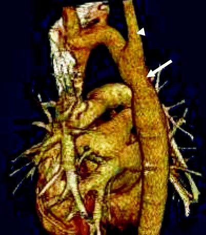
Fig. 32.1
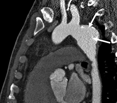
A 25-year-old man after surgical repair in early childhood of aortic coarctation using the end-to-end anastomosis technique. This oblique sagittal scan shows the normal postoperative appearance of a repaired coarctation. The arrow points to the site of the anastomosis. The arrowhead points to the origin of left subclavian artery above the site of repair. There was no significant gradient at the anastomotic site
Less frequently used techniques include insertion of a graft in situ or in an extra-anatomic location (Fig. 32.2). Interposition grafts using aortic homografts have been used when the resected segment of coarctation is too long to allow an end-to-end anastomosis. Interposition graft insertion creates proximal and distal anastomoses.
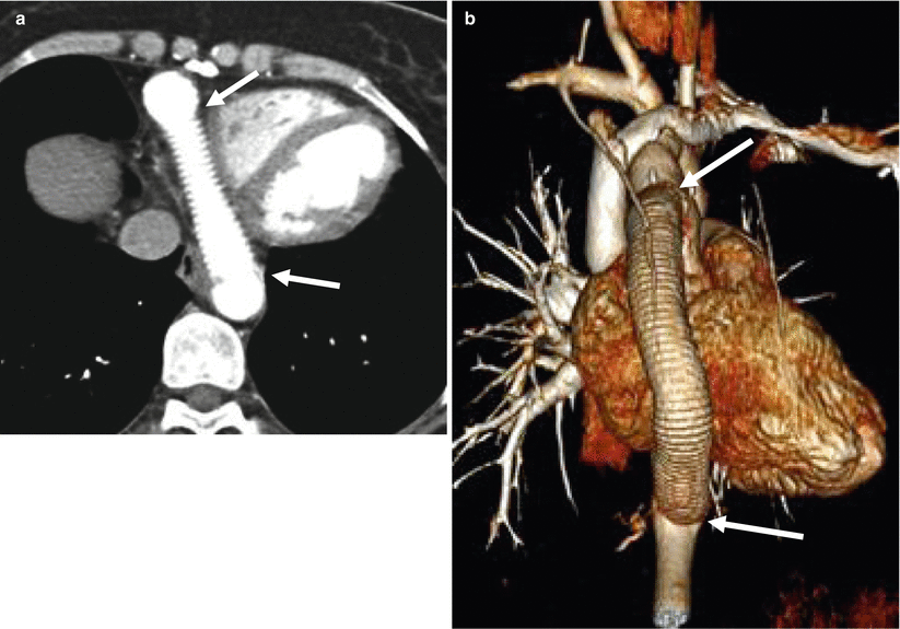
Fig. 32.2
Graft interposition. A 38-year-old woman who had a patch repair of an aortic coarctation as a child and subsequent ascending to descending aortic bypass graft placed when she had a recurrent coarctation. Panel (a) is an axial scan and panel (b) is a volume-rendered reformat showing the normal appearance of a bypass graft from the ascending to descending aorta. This procedure is used in patients with long-segment aortic coarctation. The arrows point to the proximal and distal anastomoses of the graft
32.1.2 Endovascular Repairs
Balloon Angioplasty
Percutaneous balloon angioplasty is often used in the treatment of residual stenosis or recoarctation. Its use for treatment of native coarctation is more controversial, although it has been recommended as an alternative to surgery in adolescents and adults [4, 5]. Its use is limited in younger patients because restenosis is common [6].
Stent Placement
Aortic stent placement has been used in patients with primary coarctation as well as in those with recurrent coarctation after surgical repair or balloon angioplasty (Fig. 32.3) [7]. Recurrence is a frequent problem after balloon dilatation, and endovascular stents have become an integral component for the treatment of recurrent lesions following balloon angioplasty.
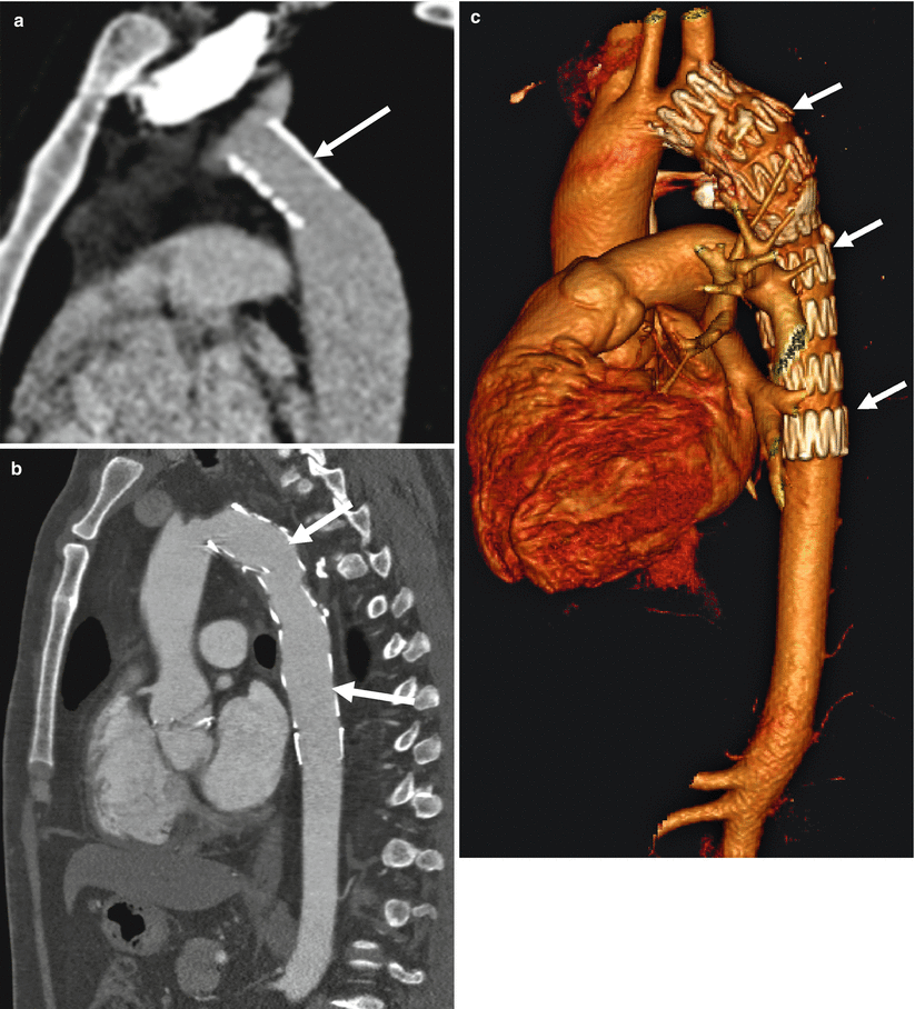
Fig. 32.3
Stent placement. Two patients with history of coarctation who had a patch repair of the coarctation at young ages and developed recurrent aortic coarctation. Panels (a), (b) and (c) are oblique sagittal views showing complete resolution of stenosis after balloon dilatation and stent placement (arrows) in the region of coarctation. No pressure gradient was seen at the end of procedure in either patient
32.2 Post-Treatment Complications
Following the use of surgical or endovascular repairs, survival has improved dramatically, although life expectancy is not that of unaffected peers. Post-intervention, morbidity and mortality are often determined by associated major cardiovascular anomalies, degree of systemic hypertension, and complications of the repair [8, 9]. The most common complications associated with surgical and endovascular techniques are recurrent coarctation and aneurysm formation.
32.2.1 Recurrent Coarctation or Restenosis
Recurrent coarctation is considered to be present whenever the pressure gradient across the anastomotic stenosis is higher than 20 mmHg at rest and occurs after both surgical and endovascular treatment (Fig. 32.4). Residual coarctation is defined as the presence of a gradient in the early postoperative period.
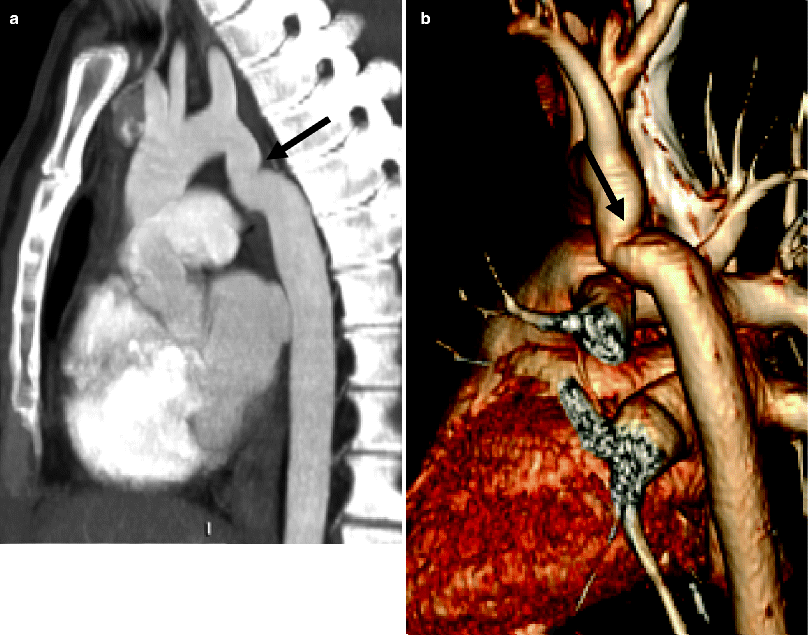
Fig. 32.4
Recurrent coarctation. A 24-year-old female with a history of coarctation of the aorta status post-balloon angioplasty 6 months previously who presented again with increasing hypertension and a recurrent gradient of 25 mmHg. Panels (a) and (b) are two sagittal computed tomographic views showing a focal tortuosity and mild narrowing of the proximal descending aorta (arrows). She subsequently underwent stenting
The risk of recurrent coarctation increases if surgical repair is performed before 1 year of age, while the risk of hypertension increases when repair is performed after 1 year of age [8, 10, 11]. In subjects who have undergone patch aortoplasty, hemodynamically significant recurrent coarctation was reported in up to 50 % of survivors who had repair in the neonatal period with an overall recurrence rate of about 5 % [12].
32.2.2 Aneurysm Formation
Aneurysm formation is defined as an area of focal dilatation with a diameter that is 1.5 times that of the aorta at the level of the diaphragm (Figs. 32.5 and 32.6). Aneurysm formation occurs after both surgical and endovascular treatment, with an incidence of about 5 % [13]. Its occurrence is most common after patch aortoplasty [12]. Approximately 7 % of deaths after coarctation repair have been attributed to aneurysm formation [8]. Delayed aneurysm formation in the ascending aorta, proximal to the surgical repair site, has also been reported as a late complication (Fig. 32.7) [14, 15].