(1)
Department of Neuroscience, University of Turin Ospedale Molinette, Turin, Italy
Abstract
In the study of the normal aortic arch and brachiocephalic arteries and of their possible variants, some short considerations about the embryogenesis are necessary.
In the study of the normal aortic arch and brachiocephalic arteries and of their possible variants, some short considerations about the embryogenesis are necessary.
An important aspect in the embryological development of the cerebrovascular system, as pointed out by Streeter (1918), is that it is not an independent process but it is linked to the progressive development of the brain to which the vascular structure continuously adapts.
The vascular structures develop from primitive vascular arches (Congdon 1922; Padget 1948; Haughton and Rosenbaum 1974). These are longitudinal vessels arising on each side from the ductus arteriosus having an ascending course forming the primitive ventral (ascending) paired aorta. The vessels then bend dorsally continuing caudally in the paired primitive descending aorta. From these arches arise the brachiocephalic arteries. In the embryogenesis, six arches in different phases develop and progressively disappear. The final normal aortic arch is characterized by the persistence of the left fourth primitive ventral arch from which arise (right to left) the brachiocephalic trunk (innominate artery), the left common carotid artery, and the left subclavian artery. From the brachiocephalic trunk arise the right common carotid artery and the subclavian artery, giving off the right vertebral artery. The left vertebral artery arises from the left subclavian artery (Figs. 1.1, 1.2, and 1.3).
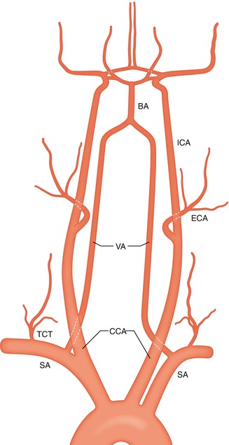
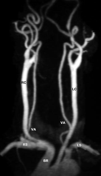
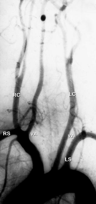
Fig. 1.1
Drawing showing the aortic arch, the extra- and intracranial cerebral arteries. Subclavian artery (SA). Thyrocervical trunk (TCT). Common carotid artery (CCA). Vertebral artery (VA). Internal carotid artery (ICA). External carotid artery (ECA). Basilar artery (BA). Circle of Willis
Fig. 1.2
Normal aortic arch, magnetic resonance imaging (MRI) angiography. Brachiocephalic trunk (BR), from which arise the right common carotid artery (RC) and right subclavian artery (RS). Common left carotid artery (LC), left subclavian artery (LS). Normal origin of both vertebral arteries (VA). The bifurcation of the two common carotid arteries is well demonstrated
Fig. 1.3
Normal aortic arch angiogram with typical origin of the left and right common carotid arteries (LC, RC). Subclavian arteries (RS, LS), asymmetry of the vertebral arteries (VA). That of the left is hypoplastic
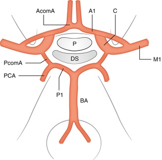
Considering the embryogenesis of the brachiocephalic arteries, from each common carotid artery arises the external carotid artery which supplies the extracranial and meningeal territories and the internal carotid artery (ICA) which divides intracranially into a cranial (anterior) and caudal (posterior) division (Padget 1944, 1948; Lazorthes 1961 Kier 1974; Lazorthes et al. 1976). From the cranial division arise progressively the anterior choroidal, the anterior cerebral, and the middle cerebral arteries responsible for the supply of the cerebral hemispheres. From the caudal division arises the posterior communicating artery (Pcom A) which gives off at its distal end the medial posterior choroidal artery and a mesencephalic–diencephalic branch from which arises the lateral posterior choroidal artery. In the further evolution, the PcomA continues in the posterior cerebral artery (PCA) which progressively extends supplying the posterior part of the cerebral hemisphere. The PcomA (pars carotica of PCA) is connected with bilateral longitudinal channels (BLC) closely placed on the surface of the primitive brainstem forming the primitive duplicated basilar artery (BA) which later fuse together in the median BA. The cranial part of these longitudinal channels will become the P1 segment (pars basilaris of the PCA). Also at this stage of the evolution, the primary ICA is connected with the BLC through transitory arteries (trigeminal, otic, hypoglossal, and proatlantal) which normally disappear (see Sect. 2.5). The flow is directed from cranial to caudal. To the proximal part of the BA converge the two vertebral arteries (VAs) formed by longitudinal anastomotic channels connected proximally with the subclavian artery. The connection of the VAs with the BA leads to an inversion of the flow which is now from caudal to cranial.
From the vertebral and basilar arteries arise the vessels supplying the brainstem and cerebellum. The cerebellar arteries are the latest to develop due to the late development of the cerebellum.
At the 6–7 weeks of the fetal development (De Vriese 1905; Padget 1944–1948), at the base of the cerebrum, both carotid and basilar arteries are connected to each other by the way of the small anastomotic circle called “circle of Willis” (Willis 1684). Both anterior cerebral arteries are linked by the anterior communicating artery, and each carotid artery is connected through the posterior communicating artery with the respective PCA (Fig. 1.4).