Anatomy and Physiology
The cardiovascular system’s job is to deliver oxygenated blood to tissues and remove waste products. The heart pumps blood to all organs and tissues of the body, and the autonomic nervous system (ANS) controls how the heart pumps.
CARDIOVASCULAR ANATOMY
Consisting of the arteries and veins, the vascular network:
carries blood throughout the body
keeps the heart filled with blood
maintains blood pressure.
Structures of the heart
The heart is a hollow, muscular organ about the size of a closed fist. It’s about 5″ (12.5 cm) long, 3½″ (9 cm) in diameter at its widest point and weighs 1 to 1¼ 1b (453.5 to 567 g). The heart is located between the lungs in the mediastinum, behind and to the left of the sternum.
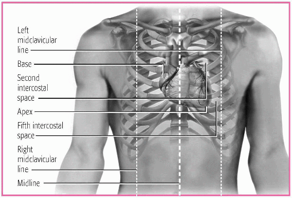
The heart spans the area from the second to the fifth intercostal space. The heart’s right border lines up with the right border of the sternum. The heart’s left border lines up with the left midclavicular line. The heart’s base, its posterior surface, is the widest part of the heart. It’s formed mainly by the left atrium, which is located at the top of the heart. The apex, formed mainly by the left ventricle, is located at the fifth left intercostal space at the bottom of the heart. The apex is the point of maximum impulse, where heart sounds are the loudest. (See Locating the heart, page 2.)
Leading into and out of the heart are the great vessels:
LOCATING THE HEART
The heart lies beneath the sternum within the mediastinum. In most people, two-thirds of the heart extends to the left of the body’s midline, close to the left midclavicular line. The heart rests obliquely so that its broad part, the base, is at its upper right and the pointed end, the apex, is at its lower left.
 |
HEART WALL
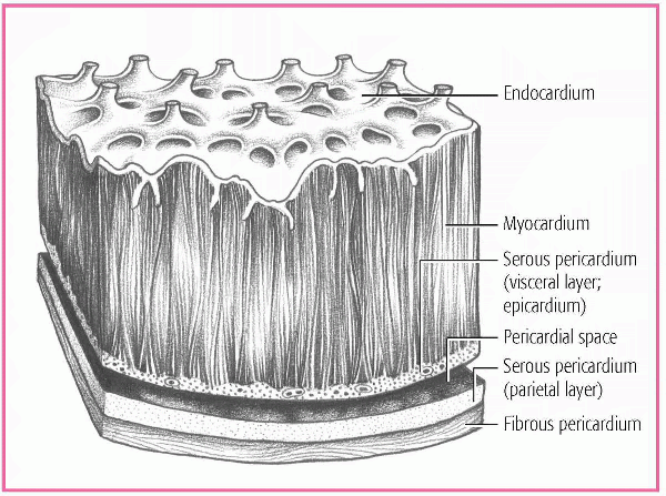
The heart wall consists of three layers:
endocardium, a thin layer of endothelial tissue
myocardium, composed of interlacing bundles of thick cardiac muscle fibers, which forms most of the heart wall
epicardium, which makes up the outside layer and is made of connective tissue covered by the epithelium.
The pericardium is a fibroserous sac that surrounds the heart and the roots of the great vessels. It consists of the serous pericardium and the fibrous pericardium. The serous pericardium contains two layers:
parietal layer, which lines the inside of the fibrous pericardium
visceral layer, which adheres to the surface of the heart.
The fibrous pericardium is a thicker layer that protects the heart. The space between the two layers, called the pericardial space, contains
10 to 30 ml of serous fluid, which prevents friction between the layers as the heart pumps. (See A look at the layers of the heart wall.)
10 to 30 ml of serous fluid, which prevents friction between the layers as the heart pumps. (See A look at the layers of the heart wall.)
HEART CHAMBERS
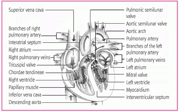
The heart contains four hollow chambers: two atria and two ventricles. The right atrium lies in front of and to the right of the smaller but thicker-walled left atrium. An interatrial septum separates the two chambers and helps them contract. The right and left atria serve as volume reservoirs for blood being sent into the ventricles. The right atrium receives deoxygenated blood returning from the body through the inferior and superior vena cavae and from the heart through the coronary sinus. The left atrium receives oxygenated blood from the lungs through the four pulmonary veins. Contraction of the atria forces blood into the ventricles. (See A look at the internal structures of the heart, page 4.)
The right and left ventricles make up the two lower chambers. The right ventricle:
lies behind the sternum
forms the largest part of the sternocostal surface and inferior border of the heart
receives deoxygenated blood from the right atrium
pumps the deoxygenated blood through the pulmonary arteries to the lungs where it’s reoxygenated.
The left ventricle:
forms the apex and most of the left border of the heart and its posterior and diaphragmatic surfaces
receives oxygenated blood from the left atrium
pumps oxygenated blood through the aorta into the systemic circulation.
The interventricular septum separates the ventricles and helps them pump.
The thickness of a chamber’s walls is determined by the amount of pressure needed to eject its blood. Because the atria act as reservoirs for the ventricles and pump the blood a shorter distance, their walls are considerably thinner than the walls of the ventricles. Likewise, the left ventricle has a much thicker wall than the right ventricle because the left ventricle pumps blood against the higher pressures in the aorta.
The right ventricle pumps blood against the lower pressures in the pulmonary circulation.
The right ventricle pumps blood against the lower pressures in the pulmonary circulation.
BLOOD FLOW THROUGH THE HEART
Deoxygenated venous blood returns to the right atrium through three vessels:
superior vena cava, which carries blood from the upper body
inferior vena cava, which carries blood from the lower body
coronary sinus, which carries blood from the heart muscle.
The increasing volume of blood in the right atrium raises the pressure in that chamber above the pressure in the right ventricle. Then the tricuspid valve opens, allowing blood to flow into the right ventricle.
The right ventricle pumps blood through the pulmonic valve into the pulmonary arteries and lungs, where oxygen is picked up and excess carbon dioxide is released. From the lungs, the oxygenated blood flows through the pulmonary veins and into the left atrium. This completes a circuit called pulmonary circulation.
As the volume of blood in the left atrium increases, the pressure in the left atrium exceeds the pressure in the left ventricle. The mitral valve opens, allowing blood to flow into the left ventricle. The ventricle contracts and ejects the blood through the aortic valve into the aorta. The blood is distributed throughout the body, releasing oxygen to the cells and picking up carbon dioxide. Blood then returns to the right atrium through the veins, completing a circuit called systemic circulation.
Coronary circulation
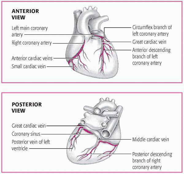
Like the brain and other organs, the heart needs an adequate supply of oxygenated blood to survive. The main coronary arteries lie on the surface of the heart, with smaller arterial branches penetrating the surface into the cardiac muscle mass. The heart receives its blood supply almost entirely through these arteries. Only a small percentage of the heart’s endocardial surface can obtain sufficient amounts of nutrition directly from the blood in the cardiac chambers. (See A look at coronary circulation, page 6.)
Understanding coronary blood flow can help you provide better care to a patient with coronary artery disease because you’ll be able to predict which areas of the heart will be affected by a narrowing or occlusion of a particular coronary artery.
The left main and right coronary arteries arise from the coronary ostia, small orifices located just above the aortic valve cusps. The right coronary artery fills the groove between the atria and ventricles, giving
rise to the acute marginal artery and ending as the posterior descending artery. The right coronary artery supplies blood to:
rise to the acute marginal artery and ending as the posterior descending artery. The right coronary artery supplies blood to:
right atrium
right ventricle
inferior wall of the left ventricle
sinoatrial (SA) node in about 50% of the population
atrioventricular (AV) node in about 90% of the population.
The posterior descending artery supplies the posterior wall of the left ventricle in most people.
The left main coronary artery varies in length from a few millimeters to a few centimeters. It splits into two major branches, the left anterior descending (also known as the interventricular) and the left circumflex
arteries. The left anterior descending artery runs down the anterior surface of the heart toward the apex. This artery and its branches—the diagonal arteries and the septal perforators—supply blood to:
arteries. The left anterior descending artery runs down the anterior surface of the heart toward the apex. This artery and its branches—the diagonal arteries and the septal perforators—supply blood to:
anterior wall of the left ventricle
anterior interventricular septum
bundle of His
right bundle branch
anterior fasciculus of the left bundle branch.
The circumflex artery circles the left ventricle, ending on its posterior surface. The obtuse marginal artery arises from the circumflex artery. The circumflex artery provides oxygenated blood to:
lateral wall of the left ventricle
left atrium
posterior wall of the left ventricle in 10% of people
posterior fasciculus of the left bundle branch
SA node in about 50% of people
AV node in about 10% of people.
In most people, the right coronary artery is the dominant vessel, meaning the right coronary artery supplies the posterior wall via the posterior descending artery. This is called right coronary dominance or a dominant right coronary artery. Likewise, when the left coronary artery supplies the posterior wall via the posterior descending artery, the terms left coronary dominance or dominant left coronary artery are used.
When two or more arteries supply the same region, they usually connect through anastomoses, junctions that provide alternative routes of blood flow. This network of smaller arteries, called collateral circulation, provides blood to capillaries that directly feed the heart muscle. Collateral circulation often becomes so strong that even if major coronary arteries become narrowed with plaque, collateral circulation can continue to supply blood to the heart.
In contrast to the other vascular beds in the body, the heart receives its blood supply primarily during ventricular relaxation or diastole, when the left ventricle is filling with blood. This is because the coronary ostia lie near the aortic valve and become partially occluded when the aortic valve opens during ventricular contraction or systole. However, when the aortic valve closes, the ostia are unobstructed, allowing blood to fill the coronary arteries. Because diastole is the time when the coronary arteries receive their blood supply, anything that shortens diastole, such as periods of increased heart rate or tachycardia, will also decrease coronary blood flow.
In addition, the left ventricle compresses intramuscular blood vessels during systole. During diastole, the cardiac muscle relaxes, and blood flow through the left ventricular capillaries is no longer obstructed.
Just like the other parts of the body, the heart has its own veins, which remove oxygen-depleted blood from the myocardium. About three quarters of the total coronary venous blood flow leaves the left ventricle by way of the coronary sinus, an enlarged vessel that returns blood to the right atrium. Most of the venous blood from the right ventricle flows directly into the right atrium through the small anterior cardiac veins, not by way of the coronary sinus. A small amount of coronary blood flows back into the heart through the thebesian veins, minute veins that empty directly into all chambers of the heart.
Heart valves
Valves in the heart keep blood flowing in only one direction through the heart, preventing blood from traveling the wrong way. Healthy valves open and close passively as a result of pressure changes within the four chambers.
Stay updated, free articles. Join our Telegram channel

Full access? Get Clinical Tree


