Anatomy and physiology
Location of the heart
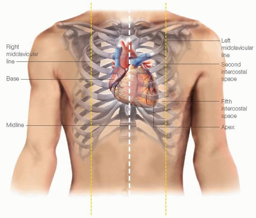
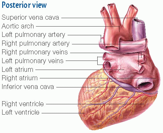
The heart lies beneath the sternum within the mediastinum, a cavity that contains the tissues and organs separating the two pleural sacs. In most people, two-thirds of the heart extends to the left of the body’s midline, close to the left midclavicular line. The heart rests obliquely so that its broad part, the base, is at its upper right and the pointed end, the apex, is at its lower left. The apex is the point of maximal impulse, where heart sounds are loudest.
|

|
Structures of the heart
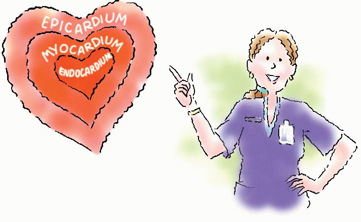
Heart wall layers
The epicardium, the outer layer, is made up of squamous epithelial cells overlying connective tissue.
The myocardium, the middle layer, forms most of the heart wall. It has striated muscle fibers that cause the heart to contract.
The endocardium, the heart’s inner layer, consists of endothelial tissue with small blood vessels and bundles of smooth muscle.
|
|
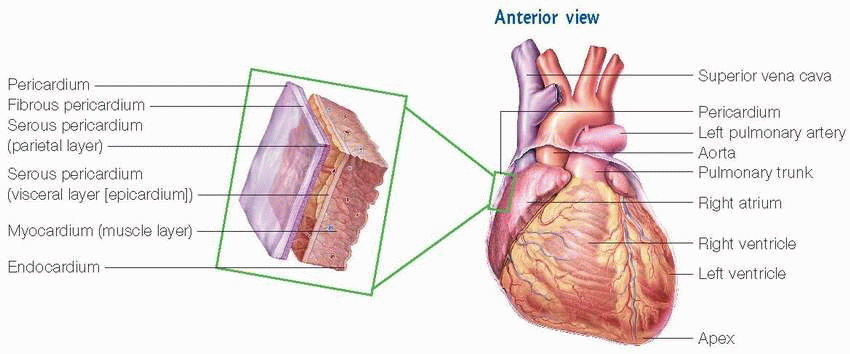
A sac called the pericardium surrounds the heart and roots of the great vessels. It consists of two layers: the fibrous pericardium (tough, white fibrous tissue) and serous pericardium (thin, smooth inner portion). The serous pericardium also has two layers: the parietal layer (lines the inside of the fibrous pericardium) and the visceral layer (adheres to the surface of the heart).
Between the fibrous and serous pericardium is the pericardial space. This space contains pericardial fluid, which lubricates the surfaces of the space and allows the heart to move easily during contraction.
|
Heart chambers
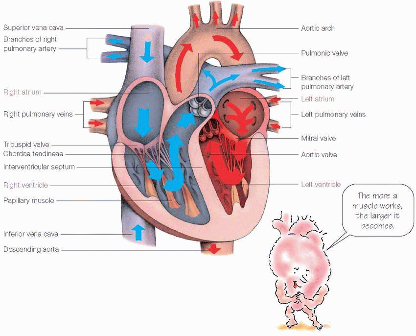
Within the heart lie four hollow chambers, two atria and two ventricles:
The right and left atria serve as volume reservoirs for blood being sent into the ventricles.
The interatrial septum divides the atrial chambers, helping them to contract and force blood into the ventricles below.
The ventricles serve as the pumping chambers of the heart.
The interventricular septum separates the ventricles and also helps them to pump.
Pump it up!
The thickness of a chamber’s wall depends on the amount of high-pressure work the chamber does:
Because the atria only have to pump blood into the ventricles, their walls are relatively thin.
The walls of the right ventricle are thicker because it pumps blood against the resistance of the pulmonary circulation.
The walls of the left ventricle are thickest of all because it pumps blood against the resistance of the systemic circulation.
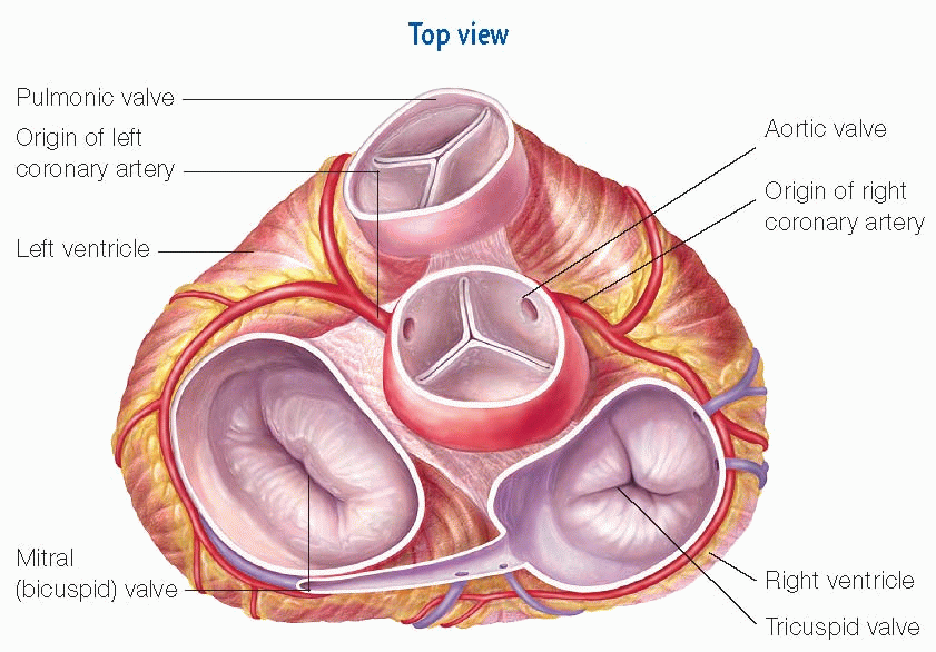
Heart valves
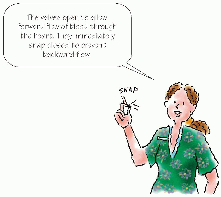
The heart contains four valves: two atrioventricular (AV) valves (the mitral and tricuspid) and two semilunar valves (the pulmonic and aortic). The valves allow forward flow of blood through the heart and prevent backward flow
The tricuspid valve, or right AV valve, prevents backflow from the right ventricle into the right atrium. The mitral valve, also known as the bicuspid or left AV valve, prevents backflow from the left ventricle into the left atrium. The pulmonic valve, one of the two semilunar valves, prevents backflow from the pulmonary artery into the right ventricle. The other semilunar valve is the aortic valve, which prevents backflow from the aorta into the left ventricle.
|
Under pressure!
Pressure changes within the heart affect the opening and closing of the valves. The amount of blood stretching the chamber and the degree of contraction of the chamber wall determine the pressure. For example, as blood fills a chamber, the pressure rises; then, as the chamber wall contracts, the pressure rises further. This increase in pressure causes the valve to open and blood to flow out into an area of lower pressure, leading to an equal pressure state.
|
Flow of blood through the heart
|
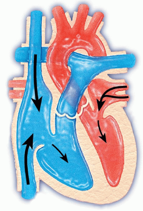
Step 1
Blood fills all heart chambers.
The right atrium receives deoxygenated blood returning from the body through the inferior and superior venae cavae and from the heart through the coronary sinus. The left atrium receives oxygenated blood from the lungs through the four pulmonary veins. Passive filling of the ventricles begins with diastole.
|
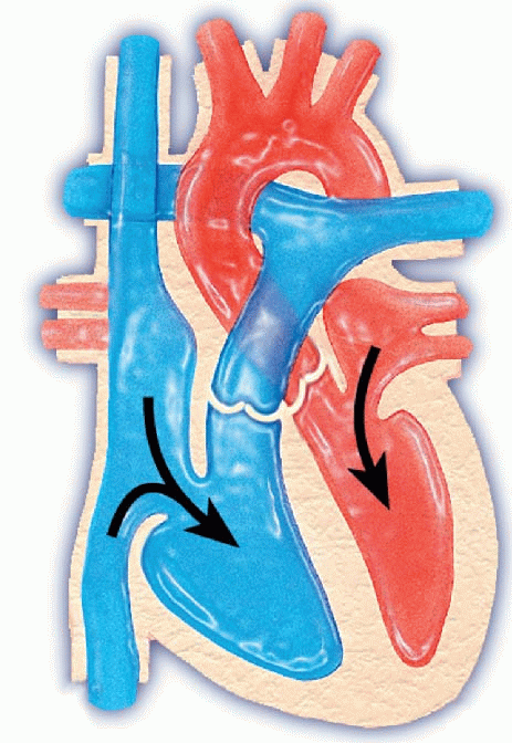
Step 2
The atria contract. The remaining blood enters the ventricles.
The atria pump their blood through the two AV valves (mitral and tricuspid) directly into their respective ventricles.
|
Stay updated, free articles. Join our Telegram channel

Full access? Get Clinical Tree


