Anatomy and physiology
The respiratory system includes the airways, lungs, bony thorax, and respiratory muscles. These structures and the central nervous system work together to deliver oxygen to the bloodstream and remove excess carbon dioxide from the body.
RESPIRATORY ANATOMY
Knowing the basic anatomy of the respiratory system will help you perform a comprehensive respiratory assessment and recognize any abnormalities. (See Structures of the respiratory system, page 2.)
Airways and lungs
The airways are divided into the upper and lower airways. The upper airways include the nasopharynx (nose), oropharynx (mouth), laryngopharynx, and larynx. Their purpose is to warm, filter, and humidify inhaled air. They also help to make sound and send air to the lower airways.
The epiglottis is a flap of tissue that closes over the top of the larynx when the patient swallows. It protects the patient from aspirating food or fluid into the lower airways. The larynx is located at the top of the trachea and houses the vocal cords. It’s the transition point between the upper and lower airways.
The lower airways begin with the trachea, which then divides into the right and left mainstem bronchial tubes. The bronchial tubes divide into bronchi, which are lined with mucus-producing ciliated epithelium, one of the lungs’ major defense systems.
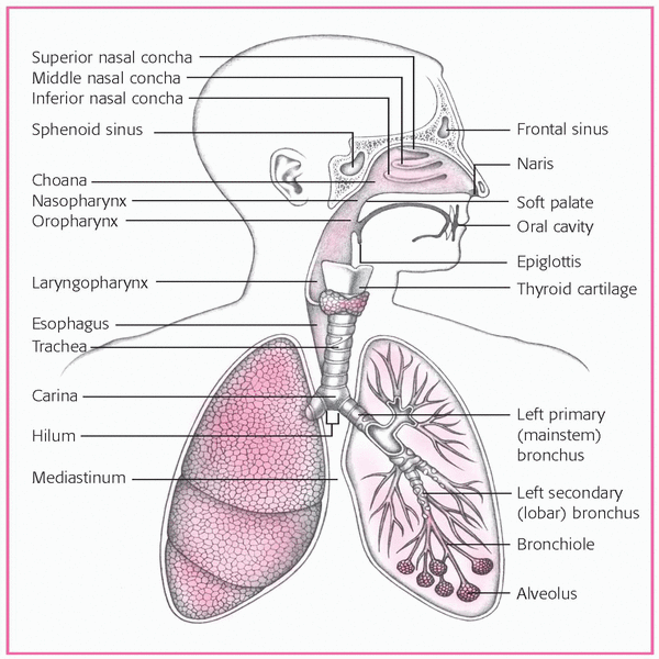
The bronchi then divide into secondary bronchi, tertiary bronchi, terminal bronchioles, respiratory bronchioles, alveolar ducts and, finally, alveoli, the gas-exchange units of the lungs. The lungs in an adult typically contain about 300 million alveoli.
Each lung is wrapped in a lining called the visceral pleura. The right lung is larger and has three lobes: upper, middle, and lower. The left lung is smaller and has only an upper and a lower
lobe. The lungs share space in the thoracic cavity with the heart and great vessels, the trachea, the esophagus, and the bronchi. All areas of the thoracic cavity that come in contact with the lungs are lined with parietal pleura.
lobe. The lungs share space in the thoracic cavity with the heart and great vessels, the trachea, the esophagus, and the bronchi. All areas of the thoracic cavity that come in contact with the lungs are lined with parietal pleura.
A small amount of fluid fills the area between the two layers of the pleura. That fluid, called pleural fluid, allows the layers of the pleura to slide smoothly over one another as the chest expands and contracts. The parietal pleura also contains nerve endings that emit pain signals when inflammation occurs.
Thorax
Composed of bone and cartilage, the thoracic cage supports and protects the lungs. The vertebral column and 12 pairs of ribs form the posterior portion of the thoracic cage. The ribs, the major portion of the thoracic cage, extend from the thoracic vertebrae toward the anterior thorax. Along with the vertebrae, they support and protect the thorax, permitting the lungs to expand and contract. The vertebrae and ribs are numbered from top to bottom. Posteriorly, certain landmarks are used to help identify specific vertebrae. In 90% of people, the seventh cervical vertebra (C7) is the most prominent vertebra on a flexed neck; for the remaining 10%, it’s the first thoracic vertebra (T1). Thus, to locate a specific vertebra, count along the vertebrae from C7 or T1. (See Respiratory assessment landmarks, page 4.)
The manubrium, sternum, xiphoid process, and ribs form the anterior thoracic cage, which has the additional duty of protecting the mediastinal organs that lie between the right and left pleural cavities. Ribs 1 through 7 attach directly to the sternum; ribs 8 through 10 attach to the cartilage of the preceding rib. The other two pairs of ribs are “free floating”; they don’t attach to any part of the anterior thoracic cage. Rib 11 ends anterolaterally and rib 12 ends laterally. The lower parts of the rib cage (the costal margins) near the xiphoid process form the borders of the costal angle—an angle of about 90 degrees normally.
Above the anterior thorax is a depression called the suprasternal notch. Because this notch isn’t covered by the rib cage like the rest of the thorax, it allows you to palpate the trachea and check aortic pulsation.
RESPIRATORY ASSESSMENT LANDMARKS
The common landmarks used in respiratory assessment are illustrated below.
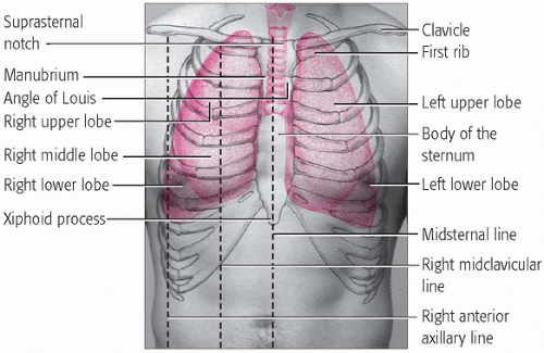 Figure. ANTERIOR VIEW |
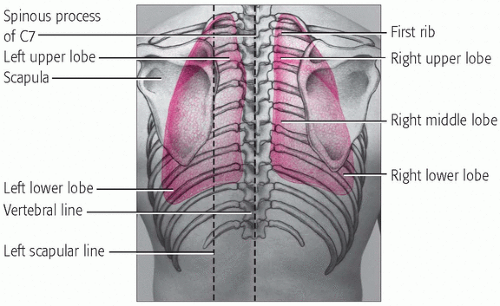 Figure. POSTERIOR VIEW |
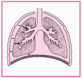
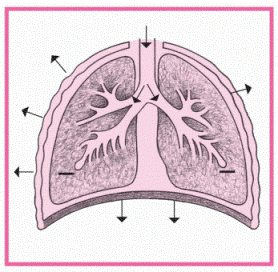
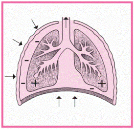
THE MECHANICS OF BREATHING
These illustrations show how mechanical forces, such as the movement of the diaphragm and intercostal muscles, produce a breath. A plus sign (+) indicates positive pressure, and a minus sign (−) indicates negative pressure.
AT REST | INSPIRATION | EXPIRATION |
|---|---|---|
|
|
|
• Inspiratory muscles relax. Stay updated, free articles. Join our Telegram channel
Full access? Get Clinical Tree
 Get Clinical Tree app for offline access

|