Acute Respiratory Distress Syndrome

Chapter 36 Acute Respiratory Distress Syndrome


The acute respiratory distress syndrome (ARDS) is an inflammatory disease process of the lungs that is a response to both direct and indirect insults, characterized clinically by severe hypoxemia, reduced lung compliance, and bilateral radiographic infiltrates. Although much has evolved in the current understanding of its pathogenesis and factors affecting patient outcome, there is still no specific treatment for ARDS.


In 1967, Ashbaugh and colleagues published an article in The Lancet in which they described for the first time a clinical syndrome that they termed the “adult respiratory distress syndrome” for its similarity to the well-recognized infant respiratory distress syndrome. From a cohort of 272 patients who were receiving respiratory support, these investigators identified 12 patients with a constellation of specific pulmonary findings: The respiratory distress in these patients was defined as sudden and catastrophic and often associated with a multiorgan system insult that led to tachypnea, hypoxemia, decreased lung compliance, and bilateral pulmonary infiltrates on the chest radiograph, in the absence of cardiogenic pulmonary edema. In this study, respiratory support consisted of oxygen therapy delivered by nasal prongs or face mask and mechanical ventilation. The mortality rate was 58%, and on histopathologic examination, the lungs of the nonsurvivors were heavier than normal and exhibited atelectasis with interstitial and alveolar edema and hyaline membranes. Since that time, the hallmarks of this syndrome, now known as ARDS, have been recognized to consist of (1) a risk factor for the development of acute respiratory distress (e.g., sepsis, trauma, pneumonia, aspiration, pancreatitis), (2) severe hypoxemia despite a relatively high fraction of inspired oxygen (FIO2), (3) decreased lung compliance, (4) bilateral pulmonary infiltrates, and (5) lack of clinical evidence of cardiogenic pulmonary edema. Acute lung injury (ALI) resulting in ARDS also can occur in the setting of left ventricular failure, but this is very difficult to diagnose without careful serial measurements of pulmonary artery and capillary (wedge) pressure. Although ALI/ARDS had been recognized for more than a century and was referenced in published data from the Second World War, it was not until the landmark paper by Ashbaugh’s group that broad clinical interest in ARDS began to emerge. In the subsequent years, very few acronyms have received as much attention in critical care medicine.


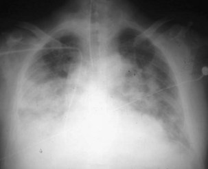
ARDS usually occurs in previously healthy people. Characteristically, there is a latent period between the insult and the development of the full-blown clinical syndrome, which usually is 18 to 24 hours in duration. After this interval, tachypnea, labored breathing, and cyanosis are observed, and arterial blood gas analysis confirms hypoxemia. The abnormalities in lung mechanics and oxygenation are assessed once the patient is intubated and receiving mechanical ventilation. The chest radiograph classically shows diffuse, bilateral, interstitial alveolar infiltrates (Figure 36-1). Resolution of the infiltrates, if it occurs at all, is much slower than with cardiogenic pulmonary edema.


Pathophysiology, Histopathology, and Etiology


ARDS is caused by an insult to the alveolar-capillary membrane that results in increased permeability and subsequent interstitial and alveolar edema. The mechanisms whereby a wide variety of insults can lead to this syndrome are not clear. ALI includes injury to both the pulmonary capillary endothelium and the alveolar epithelium. In the ARDS lung, an influx of protein-rich edema fluid into the air spaces occurs as a consequence of increased permeability of the alveolar-capillary barrier. The degree of alveolar epithelial injury is an important determinant and predictor of outcome. The normal alveolar epithelium has two types of cells. The type I cells make up 90% of the alveolar surface area and are easily injured. The type II cells make up the remaining 10% of the alveolar surface area and are more resistant to injury; their functions include surfactant production, ion transport, and proliferation and differentiation to type I cells after injury.


The loss of epithelial integrity in ARDS has several consequences. First, under normal conditions, the epithelial barrier is much less permeable than the endothelial barrier; thus epithelial injury can contribute to alveolar flooding. Second, the loss of epithelial integrity and injury to type II cells serve to disrupt normal epithelial fluid transport, impairing the removal of edema fluid from the alveolar space. Third, injury to type II cells reduces the production and turnover of surfactant. Fourth, loss of epithelial barrier can lead to sepsis in patients with bacterial pneumonia. Finally, in severe alveolar epithelium injury, pulmonary fibrosis can develop. Independent of the clinical disorders associated with ARDS (Box 36-1), it is useful to think of the pathogenesis of ARDS as a result of two different pathways: a direct insult to lung cells and an indirect insult occurring as a result of an acute systemic inflammatory response. The host’s inflammatory response to the initial direct (pulmonary) or indirect (nonpulmonary) insult is a key factor in determining the development and progression of the acute injury to the lung. Despite ongoing elucidation of the role of cellular and humoral components of the inflammatory responses in the lung, the precise sequence of events leading to lung damage is still unknown. As with any form of inflammation, ALI during ARDS represents a complex process in which multiple cellular signaling pathways can propagate or inhibit damage to the lung.


The typical histopathologic features of ARDS are collectively known as diffuse alveolar damage. The early phase of ALI—the exudative phase—is characterized by leakage of protein-rich edema fluid into the lung and inflammatory cellular alveolar infiltrates. During this phase, a cytokine storm and an array of inflammatory mediators are released into the interstitium and alveolar space, perpetuating inflammation and promoting the development of atelectasis and structural damage to the lung architecture. In addition, damage to the alveolar-capillary barrier enhances the difficulty in removing the excess of extravascular lung fluid. An important source of these inflammatory mediators is neutrophils, which play a key role in the pathogenesis and progression of ALI. Human and animal studies have demonstrated migration and activation of neutrophils in the lungs, where they cause cell damage through the production of free radicals, inflammatory cytokines, and proteases. It is well accepted, however, that a single mediator does not predominate, and that several parallel and simultaneously interacting mechanisms may be involved. Clinically, this initial phase is manifested as marked hypoxemia and reduced lung compliance. Eventually, these changes evolve to a fibroproliferative phase in which capillary thrombosis, lung fibrosis, and neovascularization take place. Most nonsurvivors of ARDS die during this phase, despite aggressive ventilatory support with high inspiratory concentrations of oxygen and positive end-expiratory pressure (PEEP). However, only a small proportion of patients with ARDS die of hypoxemia. Rather, lung injury appears to predispose patients to the development of a systemic inflammatory response that culminates in multiple system organ dysfunction. A plethora of evidence suggests that the development of multiple extrapulmonary organ dysfunction is due to alveolar epithelial-endothelial barrier disruption and the migration of cytokines produced in the lungs into the systemic circulation.


More than 50 specific conditions associated with the development of this syndrome are recognized. The risk for development of ARDS depends on the predisposing clinical condition (i.e., some events are more likely than others to progress to ARDS) but also increases with the number of predisposing factors. Sepsis, bacterial pneumonia, multiple trauma, and aspiration pneumonia are the most common predisposing factors, accounting all together for more than 70% of cases; infection is the most frequent cause. Many invading organisms can trigger host innate and acquired immune systems to initiate the inflammatory cascade. The risk for development of ARDS also depends on patient characteristics. For example, alcoholism is a predisposing factor, and new data suggest the possibility of a genetic predisposition. Overall mortality from ARDS has not decreased substantially since the publication of the 1967 report, and the current survival rate approximates to 45% in all major epidemiologic series. Sepsis-related ARDS is characterized by a higher overall disease severity, poorer recovery from lung injury, and higher mortality than non–sepsis-related ARDS. Among patients with ARDS associated with combined pulmonary and nonpulmonary sources of infection, mortality is even higher. Approximately 80% of all deaths in patients with ARDS occur within 2 to 3 weeks after the onset of the syndrome. Death traditionally has been attributed to the underlying disease, the presence of sepsis, and the failure of vital organ systems other than the lung.


Definition, Incidence, and Severity


Because it is difficult to measure changes in capillary and alveolar permeability at the bedside, diagnosis of ARDS is based on a combination of clinical, oxygenation, hemodynamic, and radiographic criteria. These criteria allow the inclusion of a highly heterogeneous group of critically ill patients, because various types of lung injury can lead to a similar pulmonary response. Despite general agreement on the overall criteria on which to base a definition of ARDS (i.e., severe hypoxemia, marked decreased of lung compliance), the specific values of these variables and the preferred conditions of measurement vary greatly among clinicians and scientists. Thus, the original description of ARDS has proved to be incapable of identifying a uniform group of patients. Several of the patients in the original report of Ashbaugh and co-workers would not be classified as having ARDS today, because fluid overload was an important etiologic factor in those cases. Some investigators have questioned whether ARDS is a distinct entity. Others have suggested that ARDS should not be considered a separate syndrome but should be seen as part of the multiple system organ dysfunction syndrome. From a clinical perspective, a strict definition of ARDS may not be required, because current management is supportive. From a therapeutic standpoint, however, a more precise definition probably is necessary, because the effects on outcome of certain ventilatory and adjunctive techniques may presumably vary depending on the degree of lung injury. In terms of prognosis, a number of investigators have examined whether various parameters of oxygenation and lung mechanics would be useful in predicting outcome. In the context of research on ARDS, a very strong argument can be made for a universal definition: It would help standardize experimental and clinical studies evaluating the natural history, incidence, pathophysiology, treatment, and outcome of ARDS. It also would help in the comparison of data among various clinical studies and centers.


A good example of the problems inherent in formulation of a definition for ARDS is the wide disparity in the literature on the incidence. Reported data in the United States suggest an occurrence rate greatly in excess of that expected from current clinical experience in Europe. The most common figure cited for the annual incidence of ARDS is 75 cases per 100,000 population. This is based on an American Lung Program Task Force of the National Heart and Lung Institute in 1972. This internal report suggested that there were about 150,000 cases per year of ARDS in the United States, a value similar to the number of all new cases of cancer. In 1988, Webster and colleagues in England estimated an incidence of 4.5 cases per 100,000 population, and in 1989, Villar and associates in Spain calculated the incidence as 3.5 new cases per 100,000 population per year. Most epidemiologic studies report an ARDS incidence ranging from 4 to 8 cases per 100,000 population per year.


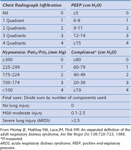
In an attempt to overcome some of these problems, Murray and colleagues proposed an expanded definition of ARDS that takes into account various pathophysiologic features of the syndrome. Their definition uses a “lung injury score” (LIS) to characterize the acute pulmonary damage by considering four components: assessment of the chest radiograph, degree of hypoxemia (determined as the ratio of arterial partial pressure of oxygen to the fraction of inspired oxygen, PaO2/FIO2), level of PEEP, and the value of lung compliance, when available (Table 36-1). The final injury score is obtained by dividing the total score by the number of components that were used. A score of 0 indicates no lung injury, a score of 1 to 2.5 indicates mild to moderate lung injury, and a score greater than 2.5 indicates severe lung injury or ARDS. The LIS is not specific for ARDS, however, and has not been validated, because it is not clear whether patients with identical LIS have similar degrees of lung injury. Furthermore, patients with a major component of cardiogenic edema may be mislabeled as having ARDS, and a postoperative patient with moderate atelectasis and mild fluid overload may fit the LIS criteria for ARDS.


Table 36-1 Lung Injury Scoring System


image

Because severe hypoxemia is the hallmark of ARDS, it should be crucial to the assessment of the severity of ARDS, for predicting the development and evolution in any given patient, and for assessing the response to treatment. To better characterize the severity of lung damage, in 1994 an American-European Consensus Conference (AECC) defined ALI and ARDS as follows:


According to these guidelines, ALI exists when the PaO2/FIO2 ratio is 300 mm Hg or less regardless of the level of PEEP and FIO2, and ARDS is present when the PaO2/FIO2 ratio is 200 mm Hg or less regardless of the PEEP setting and FIO2 (Box 36-2). Although this definition formalized the criteria for the diagnosis of ARDS and is simple to apply in the clinical setting, it has been challenged over the years in several studies. Such definitions have limitations: The physiologic thresholds do not require standardized ventilatory support, and the use of PEEP can improve oxygenation indices sufficiently to convert the patient’s status from meeting the definition of ARDS to not meeting the ARDS definition and also can change the physiology in the lung such that the patient does not meet the criteria for ARDS. Therefore, the ARDS criteria may be met when the PaO2 is measured with zero PEEP but not when measured at a PEEP of 5 or 10 cm H2O, making patient comparisons difficult. Furthermore, most of the randomized controlled studies did not use the same definition for ARDS, nor did they evaluate the same ventilatory approaches. Diversity among ARDS definitions is apparent in a large number of studies (Table 36-2).


Table 36-2 Definitions of Acute Respiratory Distress Syndrome in Several Published Reports


Published Study Criteria
Montgomery et al: Am Rev Respir Dis 132:485–489, 1985 PaO2/FIO2 <150 mm Hg
PCP <18 mm Hg
Villar et al: Am Rev Respir Dis 140:814–816, 1989 PaO2 ≤75 mm Hg on FIO2 ≥0.5
PCP <18 mm Hg
Bone et al: Chest 96:849–851, 1989 PaO2/FIO2 ≤150 mm Hg (with ZEEP)
OR PaO2/FIO2 ≤250 mm Hg with PEEP
PCP ≤18 mm Hg
Amato et al: N Engl J Med 338:347–354, 1998 Lung injury score ≥2.5 and PCP <16 mm Hg
Stewart et al: N Engl J Med 338:355–361, 1998 PaO2/FIO2 <250 mm Hg on PEEP of 5 cm H2O
Brochard et al: Am J Respir Crit Care Med 158:1831–1838, 1998 Lung injury score >2.5
Villar et al: Intensive Care Med 25:930–935, 1999 PaO2/FIO2 ≤150 mm Hg on PEEP ≥5 cm H2O
ARDSNet: N Engl J Med 342:1301–1308, 2000 AECC
Gattinoni et al: N Engl J Med 345:568–573, 2001 PAO2/FIO2 ≤200 mm Hg on PEEP ≥5 cm H2O
PCP ≤18 mm Hg
Villar et al: Crit Care Med 34:1311–1318, 2006 PAO2/FIO2 ≤200 mm Hg on PEEP ≥5 cm H2O and FIO2 ≥0.5
Meade et al: JAMA 299:637–645, 2008 PAO2/FIO2 <250 mm Hg
Mercat et al: JAMA 299:646–655, 2008 PAO2/FIO2 ≤200 mm Hg
PCP ≤18 mm Hg

AECC, American-European Consensus Conference; PCP, pulmonary capillary pressure; PEEP, positive end-expiratory pressure; ZEEP, zero PEEP.


Genetics


Critical care physicians have long recognized that some patients progress despite therapy, whereas others do better than predicted. It is now well accepted that these responses may be related to variations in the genome. Little is known, however, about the genes that are responsible for susceptibility to and outcome of ARDS. The search for genetic variants determining susceptibility and predicting outcome is still a developing field. The identification of important associations between genotype and clinical outcomes will have an impact on the development of more efficient genotype- or phenotype-guided therapies for patients with ALI or ARDS. The current understanding is that the pathogenesis of ARDS has a fundamental inflammatory component eliciting a response similar to that observed against any pathogen. In addition, common genetic risk factors with modest effects may be associated with disease susceptibility. Many studies have searched for genetic variations underlying ARDS susceptibility. Owing to the impracticality of classical genetic approximation in ARDS, association studies comparing unrelated ARDS cases with controls for genetic variants at specified locations of the human genome represent the prevailing study design for detecting such loci. The genetic variants explored usually are single base changes in the DNA, known as single-nucleotide polymorphisms (SNPs), because they are the most common variants across the genome. The genetic studies of ARDS have focused largely on candidate genes involved in the response to external stimulus and cell signal transduction, because those genes are assumed to be important in the immune response.


Extensive cross-species gene expression pattern comparisons in experimental models of ALI/ARDS have revealed that IL-6, an acute-phase response cytokine with pleiotropic effects, is highly upregulated. This finding is consistent with clinical studies indicating that IL-6 and other cytokines are released from the lungs in patients with ARDS; increased IL-6 concentrations are found in the bronchoalveolar lavage fluid and serum of these patients. IL-6 levels have been correlated with clinical outcome and implicated in the development of multiple system organ failure. A G/C SNP located at position −174 of the promoter region of the IL-6 gene has been shown to functionally affect the activity of the IL-6 gene promoter in vitro. To date, assessing the SNP variation for virtually all common variants of the gene has allowed subsequent studies to reveal a consistent picture for the association of the IL-6 gene with ALI and ARDS.


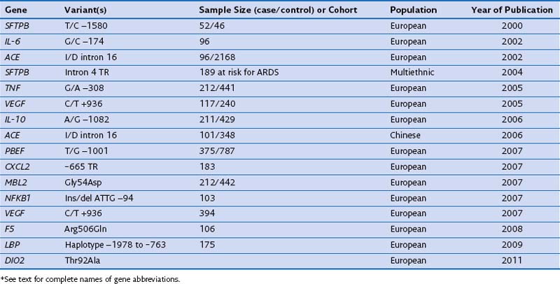
In case-control studies of patients with severe sepsis and ARDS, several investigators have explored variants of the gene encoding the lipopolysaccharide-binding protein (LBP) and serial measurements of the LBP in serum to relate them with risk gene variants. It has been reported that (1) a four-SNP risk haplotype of the LBP gene is associated with mean serum LBP concentrations within the first week of the disease process; (2) LBP levels at 48 hours are much higher in patients with ARDS than in those with ALI; and (3) a subsequent increase of LBP levels at 48 hours is associated with a four-fold increase in mortality rate. A positive association with ARDS susceptibility and/or outcome has been reported for several other genes, including surfactant pulmonary-associated protein B (SFTPB), angiotensin-converting enzyme (ACE), tumor necrosis factor (TNF), vascular endothelial growth factor (VEGF), IL-10, pre-B cell–enhancing factor (PBEF), chemokine CXC motif ligand 2 (CXCL2), mannose-binding lectin-2 (MBL2), myosin light chain kinase (MLCK), nuclear factor κ light polypeptide gene enhancer in B cells (NFKB1), coagulation factor V (F5), and type 2 deiodinase (DIO2) (Table 36-3). These genes are involved mainly in the response to external stimulus and cell signal transduction.


Altogether, significant progress has been made in the studies of genetic associations for ARDS. Because all studied candidate genes await repetitive validation in independent studies using larger samples, the search for genetic variants determining susceptibility and outcome in ARDS still needs to grow, to identify associations between genotype and clinical outcomes. The identification of genetic risk factors might allow the development of a new classification of patients and a more accurate determination of patient outcome.


Ventilator-Induced Lung Injury


Unequivocal evidence from both experimental and clinical research shows that mechanical ventilation can damage the lungs and initiate an inflammatory response, possibly contributing to extrapulmonary organ dysfunction. This type of injury, referred to as ventilator-induced lung injury (VILI), resembles the syndromes of ALI and ARDS. VILI can trigger a complex array of inflammatory mediators, resulting in a local and systemic inflammatory response. Substances produced in the lungs can be translocated into the systemic circulation as a result of injury to the pulmonary epithelium and to the capillary endothelium. This type of injury forms the basis for the use of low tidal volumes (in the range of 4 to 8 mL/kg of predicted body weight) during mechanical ventilation of patients with ALI or ARDS. The recognition of VILI has prompted a number of investigators to suggest that ALI and ARDS may in part be a product of efforts to mechanically ventilate patients, rather than representing progression of the underlying disease. On the other hand, current scientific evidence supports a link between VILI and the development of extrapulmonary organ dysfunction, in a manner similar to that in which severe cases of sepsis manifest clinically. In addition, functional genomic approaches using gene array methodology to measure lung gene expression have identified patterns of genes differentially expressed in animal models of VILI, similar to those gene pathways activated during experimental and clinical sepsis.


Ventilators are intended to deliver air or oxygen at tidal volumes and frequencies sufficient to provide adequate alveolar ventilation, to reduce the work of breathing, and to enhance oxygenation (see also Chapter 32). However, mechanical ventilation is a nonphysiologic process, and complications are associated with its application, including increased risk for pneumonia, impaired cardiac performance, and lung injury. During mechanical ventilation, pressures, gas volumes, ventilatory rates, and concentrations of inspired oxygen often are applied at levels that exceed those normally experienced by healthy lungs during spontaneous breathing. VILI is not a new concept—it is the designation historically applied to macroscopic injuries associated with alveolar rupture due to overdistention resulting from application of high inspiratory pressures. Clinical manifestations include interstitial emphysema, pneumothorax, pneumomediastinum, and pneumoperitoneum. The concept of VILI has shifted somewhat from pressure-induced (really volume-induced) injury to increased vascular permeability, accumulation of lung fluid, “atelectrauma,” and inflammation induced by mechanical ventilation.


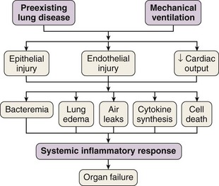
In 1998 Tremblay and Slutsky coined the term biotrauma to describe the pulmonary and systemic inflammatory response triggered by lung cell distention, alveolar disruption, and/or necrosis after the application of mechanical ventilation. Although a Consensus Conference in 1994 recommended that plateau pressure generally should be limited to 35 cm H2O, little change in ventilator practice occurred until publication of an ARDS Network study demonstrating that a lung-protective strategy using a tidal volume of 6 mL/kg of predicted body weight and moderate levels of PEEP decreased mortality in patients with ALI. This study confirmed that VILI was not just an interesting experimental entity but also was an important clinical entity. This recognition led to the widespread, albeit not universal, use of lung-protective strategies in patients with ALI. Unequivocal evidence from both experimental and clinical data has proved that mechanical ventilation can cause or aggravate ALI. Many of the pathophysiologic consequences of VILI mimic those of ARDS. A number of specific forms of injury caused by the trauma of mechanical ventilation have been identified: barotrauma, volutrauma, atelectrauma, and biotrauma. Current experimental and clinical evidence supports a link between VILI and the development of extrapulmonary organ dysfunction, by mechanisms similar to those whereby most severe cases of sepsis are clinically manifested (Figure 36-2).


Jun 12, 2016 | Posted by in RESPIRATORY | Comments Off on Acute Respiratory Distress Syndrome

Full access? Get Clinical Tree

Get Clinical Tree app for offline access