Acute Myocardial Infarction

4.4 Acute Myocardial Infarction


Acute Myocardial Infarction


Anatomical pathology:


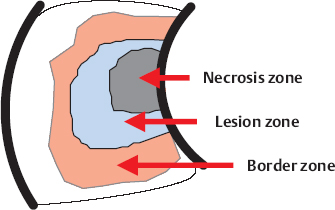
  • Necrosis zone: Electrically inactive zone (infarction Q)
  • Lesion zone: Cells markedly damaged by ischemia form abnormal potentials without participating in excitation, damaged site from which current arises — represented by ST elevation
  • Border zone: Cells participate in excitation with delayed repolarization (negative T wave)

Diagnosis:


  • Clinical, ECG changes, enzyme profile (creatine phosphokinase, troponin)
  • If two out of three criteria are positive, then infarct is confirmed


image


Definition:


  • Acute myocardial necrosis as a result of interruption of coronary perfusion
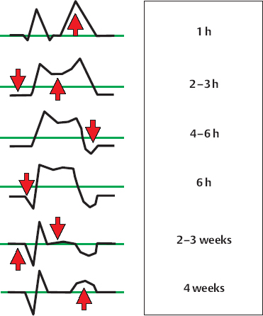
  • STEMI (ST-Elevation Myocardial Infarction): ST-elevation at least in two limb leads ≥ 0.1 mV or in two precordial leads ≥ 0.2 mV or LBBB with typical symptoms

Etiology:


  • Coronary heart disease
  • Inflammatory, trauma, spasm, embolism

Complications:


  • Bradycardia, ventricular arrhythmias
  • Aneurysm, shock, papillary muscle rupture
  • Rupture of the wall, ventricular septal defect
  • Pericarditis, Dressler syndrome


Acute treatment:


  • Reperfusion: lysis/PCTA/heparin/bypass
  • Adjuvant: nitrate, beta-blocker, sedation, oxygen
  • Treatment of complications

Chronic treatment:


  • Beta-blocker, thrombocyte aggregation inhibitor
  • CSE- and ACE-inhibitors
  • Treatment of cardiac insufficiency and arrhythmia

Myocardial Infarction: Stages and ECG Changes


Acute stages:


  1. Steeplelike/tented T waves (delayed repolarization of the inner layer as a result of acute ischemia) —early stage
  2. Depression of the isoelectric line and elevation of the ST segment (diastolic and systolic current arising from damage)—transmural ischemia
  3. Inversion of T wave (delayed repolarization) —intermediate stage
  4. Formation of an “infarction Q” (myocardial necrosis)—intermediate stage

Chronic stages:


  1. Normalization of the ST segment (following stage)
  2. Normalization of the T wave (chronic)


image

“Infarction Q Wave”


The infarcted tissue is electrically passive and forms a so-called electric hole.


The electrical vector moves forward from the infarct; a negative deflection arises in the form of a Q wave.


image

Changes in the ST Segment in Infarction


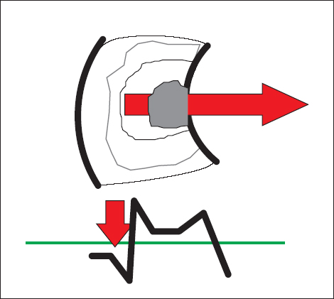
Zone of cell damage (injury) with abnormal resting potential. In diastole the cells are more electropositive than the healthy myocardium, causing the flow of current to the damaged zone with depression of the isoelectric line. In systole normal depolarization of the healthy myocardium, reversal of the flow of current to the healthy myocardium with ST elevation.

Stay updated, free articles. Join our Telegram channel

Tags:
Aug 29, 2016 | Posted by in CARDIOLOGY | Comments Off on Acute Myocardial Infarction

Full access? Get Clinical Tree

Get Clinical Tree app for offline access