of projections are required. This speed reduces breathing motion artifacts that occur when scanner rotation periods are more than 1 s.
There are two basic modes of CT scanning. By far, the most common is helical CT (the subject of Chap. 2), whereby the CT scanner rotates as the couch moves with a uniform speed. The radiation beam central axis traces a helix through the patient as the projection data are acquired. This method is the most efficient method for acquiring CT data. The couch can be moved more than the width of the collimated beam during each rotation (pitch ![$$>$$” src=”/wp-content/uploads/2016/07/A217865_1_En_3_Chapter_IEq2.gif”></SPAN>1) which, when coupled with a fast rotation rate and large longitudinal coverage, makes helical scanning extremely fast; CT scans of the thorax can be acquired within two seconds with modern scanners.</DIV><br />
<DIV class=Para>The couch motion for helical scanning is typically constrained to be constant; once the parameters of pitch, field size, and rotation rate are selected, the couch speed is fixed. While this is not a problem for standard scanning, it provides a significant constraint for 4D imaging. In order to acquire useful breathing modeling data, CT data need to be acquired during at least one breath. Breathing periods are approximately 5 s, so the minimum time that any one region of tissue needs to be imaged is 5 s. While the CT scanner rotation rate can be slowed down to guarantee that each portion of the patient is being imaged for 5 s, such slow acquisition will lead to breathing motion artifacts. The CT scan rotation period needs to be less than 0.5 s to avoid these artifacts. Rather than slow rotation rate, most helical CT scanning protocols reduce the pitch, slowing the couch and acquiring multiple images at each location within the patient. The protocols are designed such that each portion of the patient is scanned for a period of at least 5 s [<CITE><A href=]() 3, 7].
3, 7].
While helical scanning is the dominant acquisition mode and a 4D helical scanning solution has been found and implemented for most commercial systems, helical scanning has some significant limitations. First, the minimum pitch for CT scanners is approximately 0.15, which when combined with a rotation rate of 0.5 s, leads to a total amount of time that any one location is scanned of 0.5/0.06 s = 8.33 s. This amount of time is greater than the breathing period for most patients, which means that for most patients and most times, CT images will be scanned throughout the breathing cycle for the whole patient. Many patients have irregular breathing cycles and some take pauses in their breathing. The CT scanner cannot pause a helical CT acquisition, so image artifacts will occur if the patient breathing pauses for more than a few seconds. Figure 3.1 shows examples from Lu et al. [6] of breathing patterns that have such irregularities.
3.2 Benefits of Ciné CT
Ciné acquisition, in contrast to helical acquisition, is conducted with a stationary couch. The CT scanner rotates and acquires projection data over a longitudinal span that nearly matches the radiation field length. The length of this span depends on the detector layout and is usually between 2 and 4 cm (see Sect. 2.2.2). The number of acquisitions per couch position is programmed in advance and the CT rotation time determines the temporal resolution of the scan. The number of scans multiplied by the scan period is the minimum amount of time that scans can be acquired for, but pauses between scans can be programmed to lengthen the effective scan time to assure that at least one breathing period occurs at each couch position. In fact, the amount of time can easily be stretched to multiple breathing periods to reduce the chance that no motion data are acquired because of breathing irregularities.
Ciné CT acquisition provides many benefits over helical acquisition. Principally, the number of scans is selectable and not constrained by couch speed limitations. Prospective gating is also possible with ciné-CT [4]. Ciné-CT protocols can be programmed to be started by an external trigger, set for example to specific phases of the breathing cycle. Sophisticated analysis of the breathing cycle could be conducted to assure that only images that will provide the required breathing motion data will be acquired, reducing the radiation dose to the patient and improving the usability of the CT scan data.
CT images are reconstructed using x-ray projections from up to  . In helical multislice CT, the projection angles also include an out-of-plane angle. Ideally these projections would be acquired over a limited period of time. In order to minimize image noise, however, when reconstructing an image at a specific breathing phase, manufacturers utilize each projection acquired during a time consistent with that breathing phase. The projections may therefore be acquired during different breaths. If the user wants to use the commercial sorting algorithm to reconstruct images, there is no problem, but if the user wants to control the specific timepoints from which projection data are taken, this process is not useful. An advantage of ciné CT is that the projections used to acquire the CT data are acquired only during the gantry rotation time for the specific ciné acquisition. This allows the user to precisely control the timepoints at which the data are acquired for subsequent reconstructions. Pat et al. [7] compared data sufficiency conditions for helical and ciné acquisitions using a 20 cm coverage, 4 s breathing cycle, half-scan reconstruction. He found that the helical acquisition benefited from more rapid scan time (by 10 %) than the ciné acquisition. The helical slice sensitivity profile was 1.8 times broader than the ciné profile and required an additional breathing cycle of scanning at the beginning and end of the scan to assure adequate sampling at the ends of the scans. The ciné scans also delivered between 4 and 8 % less dose than the helical acquisition.
. In helical multislice CT, the projection angles also include an out-of-plane angle. Ideally these projections would be acquired over a limited period of time. In order to minimize image noise, however, when reconstructing an image at a specific breathing phase, manufacturers utilize each projection acquired during a time consistent with that breathing phase. The projections may therefore be acquired during different breaths. If the user wants to use the commercial sorting algorithm to reconstruct images, there is no problem, but if the user wants to control the specific timepoints from which projection data are taken, this process is not useful. An advantage of ciné CT is that the projections used to acquire the CT data are acquired only during the gantry rotation time for the specific ciné acquisition. This allows the user to precisely control the timepoints at which the data are acquired for subsequent reconstructions. Pat et al. [7] compared data sufficiency conditions for helical and ciné acquisitions using a 20 cm coverage, 4 s breathing cycle, half-scan reconstruction. He found that the helical acquisition benefited from more rapid scan time (by 10 %) than the ciné acquisition. The helical slice sensitivity profile was 1.8 times broader than the ciné profile and required an additional breathing cycle of scanning at the beginning and end of the scan to assure adequate sampling at the ends of the scans. The ciné scans also delivered between 4 and 8 % less dose than the helical acquisition.Fig. 3.1
Example of irregular breathing waveforms highlighting that breathing patterns can pause for times approaching 10 s. Figure is from Lu et al. [6]
Table 3.1
Summary of the comparison between helical and ciné acquisition
Ciné | Helical | |
|---|---|---|
Temporal resolution | ++ | + |
Temporal control (knowing when image data were acquired) | ++ | – |
Spatial resolution (blurring) | ++ | + |
Ability to prospectively gate | ++ | – |
Image artifacts | – | + |
Ability to manage irregular breathing | + | – |
Ability to do amplitude based sorting | ++ | ++ |
Acquisition flexibility | ++ | – |
Cone beam artifacts (assuming fan-beam ciné reconstruction) | – | ++ |
Table 3.1 shows a summary of the comparison between helical and ciné acquisitions.
3.3 Development of Volumetric CT Using Ciné CT Scans
Ciné CT scan acquisition yields a series of CT slice scans (collections), each acquired at different times and breathing phases. Between 10 and 25 repeated CT scans are acquired per table position before the table is moved to the next position, and this process is repeated until the entire volume of interest is scanned. The process of generating useful 4D CT images that encompass the organs of interest involves the building of 3D CT images at different breathing phases using the acquired ciné images.
Fig. 3.2
Process for building a 3D CT scan from sequentially acquired ciné CT image datasets. In each couch position, a number of ciné images are acquired. The image is from Low et al. [5]
Fig. 3.3
Phase-based reconstruction of 4D CT scans using a commercial phase-based sequence. The figure is from Pan et al. [8]
There are two current approaches to generate 4D CT images from ciné CT acquisitions and a breathing surrogate measured at acquisition time. They are termed amplitude-based sorting and phase-based sorting. Figure 3.2 shows an example of the amplitude-based process as described by Low et al. [5]. They selected the tidal volume measured with spirometry as the method for describing the breathing cycle. To create a 3D CT image at a specific tidal volume, they selected the ciné scans that were obtained at each couch position with a tidal volume closest to the desired volume. The result was a full 3D CT that represented the patient geometry as though the patient had been scanned at that tidal volume. This process could be repeated for any tidal volume for which there was sufficient scan data.
General Electric used a phase-based approach with their commercial ciné 4D CT scanners [8, 10]. Figure 3.3 shows the process that GE uses to acquire their 4D CT scans. The scans are acquired at the same time as a breathing surrogate is measured. The breathing cycles are divided in phases that are equal in time between inhalations. The images acquired in each phase are collected for each table position to form single-phase CT images, similar to what is done in Fig. 3.2 for breathing amplitude gating.
3.3.1 Amplitude and Phase-Angle Sorting
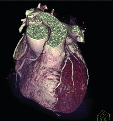
CT manufacturers have long provided robust cardiac gating acquisition and sorting tools. This is because the cardiac cycle is straightforwardly measurable and temporally stable. Typically, CT scans required well monitoring the cardiac cycle and the x-ray projections are resorted according to the cardiac phase, which is defined as the relative time between equivalent points in the cardiac cycle. Because of the stability of the cardiac cycle, this approach has been extremely successful. Figure 3.4 shows an outstanding surface rendered image of the heart from Wijesekera et al. [11]. In contrast, Fig. 3.5 shows an example of retrospectively phase-based sorted 4D CT acquired using a ciné protocol [4]. Artifacts are present at the dome of the diaphragm and tumor volume and shape (below the cross-hairs in each of the four images) are altered from phase to phase making it difficult to delineate correctly. Such artifacts are common because breathing is less reproducible than cardiac motion, and both approaches to sorting respiratory-gated CT scans are affected from this motion variability.
 < div class='tao-gold-member'>


Only gold members can continue reading. Log In or Register to continue
Stay updated, free articles. Join our Telegram channel
Full access? Get Clinical Tree
