Fig. 18.1
Broad-complex tachycardia at presentation to the local emergency department
The doctors in the Emergency Department identified a ‘broad-complex tachycardia’. Perhaps in view of the patient’s age and the absence of a history of congenital heart disease, they felt that this was most likely to be a form of supraventricular tachycardia (SVT) with aberrant conduction accounting for the broad QRS complexes. With this in mind, he was treated with intravenous adenosine at a dose of 10 mg. It is reported that the tachycardia reverted to sinus rhythm shortly after the administration of adenosine; however, there was no contemporaneous ECG recording during the administration of adenosine and precise timings were uncertain.
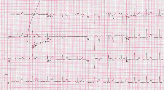
The ECG following cardioversion is shown in Fig. 18.2.
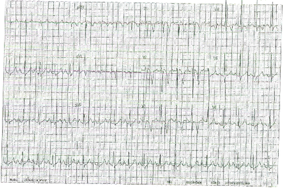
Fig. 18.2
ECG following reversion to sinus rhythm
The doctors in the Emergency Department made contact with their local Paediatric Cardiology service. The 12-lead ECGs were then reviewed with the Paediatric Electrophysiology team, who felt that the broad-complex tachycardia was likely to be a ventricular tachycardia (VT). The QRS complexes are of left bundle branch block morphology, suggesting a right ventricular origin, and the QRS axis is directed superiorly (positive in lead aVL and negative in leads II, III, and aVF), suggesting an apical origin. That the tachycardia cardioverted to sinus rhythm following adenosine administration sparked debate, as the ECG demonstrating broad-complex tachycardia fulfilled many of the conventional criteria applied for identifying VT as well as those criteria for identifying SVT with aberrancy. In terms of VT, features such as ventriculo-atrial dissociation, capture beats, and fusion beats were not obviously present. The absence of a contemporaneous ECG during adenosine administration made it impossible to understand the mode by which tachycardia was terminated, which may have permitted a more confident diagnosis of the tachyarrhythmia. Thus it remained equivocal as to whether adenosine terminated the arrhythmia or whether cardioversion occurred spontaneously, shortly after adenosine was administered.
The 12-lead ECG following termination of tachycardia was of particular interest. Sinus rhythm is clearly demonstrated and there is no manifest pre-excitation. The QRS duration is normal with no bundle branch block at baseline (therefore, any aberrancy displayed during tachycardia would have to have been rate-related) and with age-appropriate R-wave progression, which has an adult pattern in this instance. The pattern of repolarisation, however, is clearly abnormal. Though a negative T-wave in lead V1 is acceptable at this age, the deep negative T-waves in leads V1 to V4, in the absence of bundle branch block, is abnormal in a Caucasian without early repolarisation. In particular, evidence of VT of right ventricular origin in the context of a resting ECG with inverted T-waves in the anterior chest leads raises specific concerns about arrhythmogenic right ventricular cardiomyopathy (ARVC), and warrants further investigation.
Arrhythmogenic Right Ventricular Cardiomyopathy (ARVC)
Inherited cardiomyopathy characterised by:
Fibro-fatty replacement of myocytes, predominantly affecting the right ventricle
Ventricular arrhythmias and risk of sudden cardiac death
Congestive cardiac failure
Prevalence of 1:2500–1:5000 (compared to 1:500 adults for hypertrophic cardiomyopathy)
Associated with mutations in genes encoding desmosomal proteins (plakoglobin (JUP), desmoplakin (DSP), plakophilin-2 (PKP2), desmoglein-2 (DSG2), desmocollin-2 (DSC2), transforming growth factor-ß3 (TGFß3) and TMEM-43
Diagnosis based on criteria proposed by International Task Force of the Working Group of Myocardial and Pericardial Disease of the European Society of Cardiology – includes:
Evidence of fibro-fatty replacement of myocardial tissue – detected via imaging (echocardiography and cardiac MRI) and histology
ECG evidence of depolarisation and repolarisation abnormalities
Arrhythmia of right ventricular origin (though LV disease exists in ~30 %)
Positive family history including presence of known pathogenic mutations
ECG changes include:
Inverted T-waves in leads V1-V3 in the absence of RBBB (in those aged >12 years)
QRS prolongation (>110 ms) in leads V1-V3
Terminal slurring of QRS complexes in V1-V3 (≥55 ms from S-wave nadir to isoelectric line)
Epsilon waves – low amplitude deflection between QRS and T-wave
Poor R-wave progression in right praecordial leads
Low-voltage QRS complexes in the limb leads ± praecordial leads
Overall mortality rates are estimated to be 1–3 %/year and are associated with sudden cardiac death (1–2 %/year) and progressive cardiac failure
Risk factors for sudden cardiac death include: previous cardiac arrest, VT with haemodynamic compromise, significant RV dilatation/dysfunction, presence of LV disease
Having revisited the history of his previous episodes when aged 13 years, the concern was that these earlier events may have represented self-limiting runs of VT. When his 12-lead ECG from 2 years ago was reviewed (Fig. 18.3), it was noted to be very similar in appearance to his current resting ECG with evidence of markedly abnormal repolarisation, manifest as deeply negative T-waves in leads V1 to V3.
 < div class='tao-gold-member'>


Only gold members can continue reading. Log In or Register to continue
Stay updated, free articles. Join our Telegram channel
Full access? Get Clinical Tree
