Figure 87.2 Magnetic resonance images revealed (a) FSE black blood sequence images show no fat suppression. (b) Sequence images show the right ventricle apical trabeculations with maximal diastolic ratio of noncompaction (red tracing) to compacted thickness (green tracing) of >3.0.
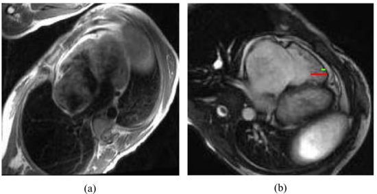
The magnetic resonance images are shown in Figure 87.2.
Management
Based on the test results and clinical manifestation, a diagnosis of RV noncompaction was made. Diuretics were administered in order to relieve the patient’s right heart failure.
Discussion
Stay updated, free articles. Join our Telegram channel

Full access? Get Clinical Tree


