6: Distal Vessel Thromboendarterectomy— How Far Can We Go?


Figure 6.1

Preoperative chest radiograph showing a prominent right atrial shadow, with central PA enlargement


Ventilation perfusion scan showed perfusion defects to the apical posterior segments of the left upper lobe, superior lingula, and the anterobasal and lateral basal segments of the left lower lobe. In the right lung there are defects in the right upper lobe, right middle lobe and basal segments of the right lower lobe (Fig. 6.2).

../images/1_1_En_6_Chapter/1_1_En_6_Fig2_HTML.jpg

Figure 6.2

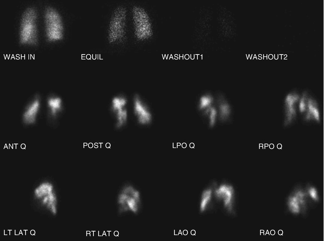
Preoperative lung scintigraphy demonstrating bilateral mismatched perfusion defects


An echocardiogram revealed RA and RV severely enlarged with depressed RV systolic function. LV size is small, with normal systolic function, TR velocity 4.5 m/s with PA systolic pressure 82 mmHg plus CVP, cardiac output 4.4 L/min, mild tricuspid regurgitation, absent pericardial effusion.


Laboratory data was as follows: Sodium 142, potassium 4.0, chloride 103, bicarbonate 24, BUN 15, creatinine 1.14, blood glucose 99. Liver function tests within normal limits except total bilirubin mildly up at 1.54. Urinalysis was negative for protein. White blood cell count 6.9, hemoglobin 13.6, hematocrit 41.5, platelets 259,000. INR 1.3, PTT 39.6.


Right heart catheterization showed right atrial mean pressure 10 mmHg, PA pressure 101/28 with a mean of 58 mmHg, Pulmonary capillary wedge pressure 13 mmHg, Cardiac output 3.30 L/min, cardiac index 1.95 L/min/m2, Pulmonary vascular resistance 1091 dynes/sec/cm-5.
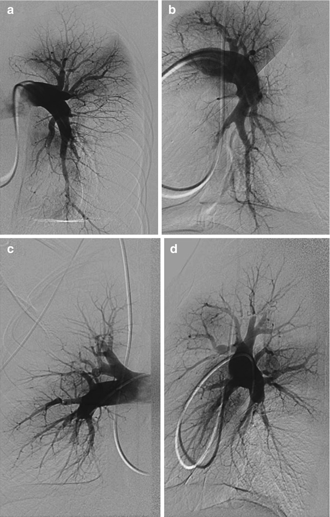

Pulmonary angiogram on the right side demonstrated a “pouch defect” in the right lower lobe; webs in the right upper lobe apical and anterior vessels. On the left side, the anterior medial vessels to the lower lobe were narrowed at the distal segmental and subsegmental level with a reduction in vascularity to the posterior LLL. Otherwise, there was a “pouch defect” in the superior segment, proximal occlusion of the lingula and a web in posterior left upper lobe (Fig. 6.3).

../images/1_1_En_6_Chapter/1_1_En_6_Fig3_HTML.jpg

Figure 6.3

(ad) Frontal (panel a) and lateral (panel b) views of the left pulmonary artery . Frontal (panel c) and lateral (panel d) views of the right pulmonary artery


She was ultimately determined to have operable CTEPH and underwent pulmonary thromboendarterectomy with a total circulatory arrest time of 67 min; 35 min for the right side and 32 min for the left. Postoperative hemodynamics were CVP 12, PAP 47/18 (28), mean 19 mmHg, CO 5.30 L/min, CI 3.2 L/min/m2 and estimated PVR 256 dynes/s/cm-5 (Fig. 6.4).

../images/1_1_En_6_Chapter/1_1_En_6_Fig4_HTML.jpg

Only gold members can continue reading. Log In or Register to continue

Stay updated, free articles. Join our Telegram channel

Oct 30, 2020 | Posted by in Uncategorized | Comments Off on 6: Distal Vessel Thromboendarterectomy— How Far Can We Go?

Full access? Get Clinical Tree

Get Clinical Tree app for offline access