CASE 59 Stenosis in a Superficial Femoral Artery
Case presentation
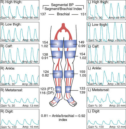
A 74-year-old diabetic man with hypertension and prior tobacco use, whose medications included aspirin, a statin, and an ACE inhibitor, developed bilateral calf claudication. Eventually, his leg symptoms progressed and he was unable to exercise, greatly limiting his lifestyle. His physician detected diminished pulses distally and assessed these symptoms with noninvasive studies (Figure 59-1). These demonstrated moderate bilateral arterial insufficiency at the level of the superficial femoral artery (SFA). He was referred for angiography.
Angiography
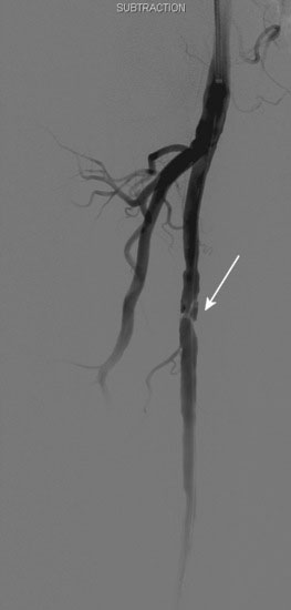
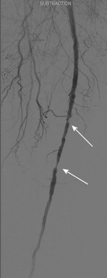
The patient’s symptoms were worse in the left leg; thus angiography of the left leg was performed first from an arterial sheath placed in the right femoral artery. It showed high-grade, sequential lesions in the mid-SFA (not shown). These were successfully treated with a self-expanding stent. Following that procedure, diagnostic angiography of the right leg was performed and demonstrated two high-grade lesions in the SFA with an ulcerated stenosis of the proximal segment of the right SFA (Figures 59-2, 59-3) and a longer, severe stenosis in the mid- to distal segment (Figure 59-4). The patient was discharged after this procedure on the left leg and experienced resolution of his left leg claudication with normalization of his left leg noninvasive studies. However, he continued to be limited by right leg claudication. Therefore, approximately 3 months after his left leg intervention, he presented for endovascular treatment of the right leg.
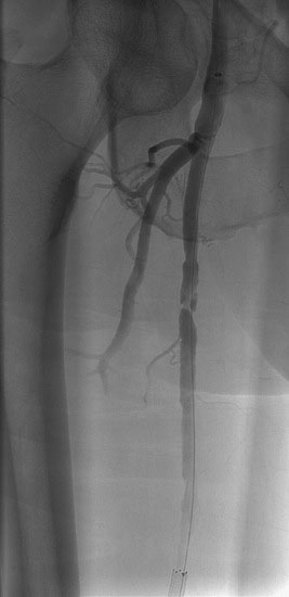
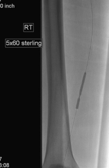
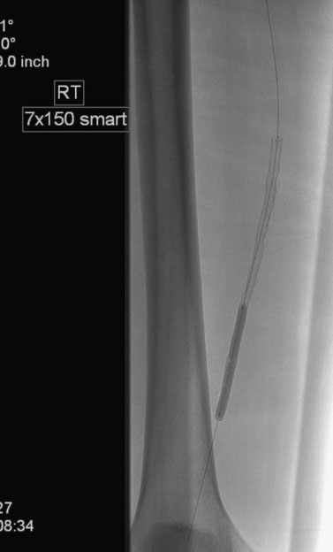
Access was obtained from the left common femoral artery. An Omniflush catheter was used to lay out the iliac vessels, and a 6 French long sheath was placed “up and over” from the left femoral to the right external iliac artery. An intravenous bolus of unfractionated heparin was administered to achieve a therapeutic activated clotting time of more than 250 seconds. The proximal and distal lesions were crossed with a hydrophilic glide wire. A catheter was then used to exchange the glide wire for a 0.018 inch guidewire. The distal SFA was treated first using a 5 mm diameter by 60 mm long balloon in two overlapping locations. The distal lesion was found to be difficult to expand (Figure 59-5); ultimately, the balloon appeared fully expanded at 12 atmospheres of pressure. A post balloon angiogram showed residual stenosis and elastic recoil; thus the operator deployed a 7 mm diameter by 150 mm long self-expanding nitinol stent across the extent of the distal lesions. The stent was postdilated with the 5 mm diameter by 60 mm long balloon (Figure 59-6) and the post-stent angiogram demonstrated an excellent result (Figure 59-7).