CASE 46 Balloon Pericardial Window
Case presentation
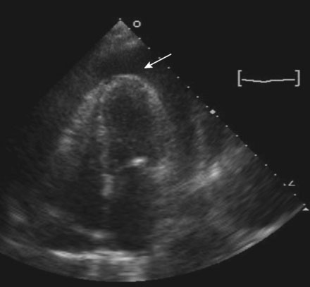
Upon presentation, she was in mild distress from dyspnea, but remained hemodynamically stable with a blood pressure of 150/70 mmHg and, although physical examination confirmed elevation of the jugular venous pressure, there was no pathologic pulsus paradoxus. The electrocardiogram showed sinus tachycardia with low voltage and echocardiography revealed a large circumferential pericardial effusion (Figure 46-1) along with right ventricular collapse and right atrial inversion consistent with tamponade. She underwent pericardiocentesis with removal of 750 mL of serosanguineous fluid. Cytological analysis of the fluid subsequently confirmed adenocarcinoma. A pericardial drain remained in place and she was admitted for continued observation. Over the ensuing 2 days, more than 200 mL of fluid drained from the pericardial space each day. She was referred to the cardiac catheterization laboratory for a balloon pericardial window.


