CASE 41 Postoperative Acute STEMI
Cardiac catheterization
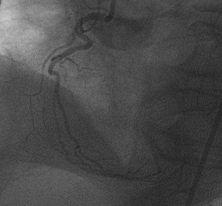
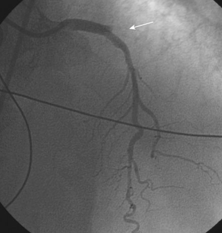
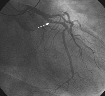
The right coronary artery was small and nondominant, with luminal irregularities (Figure 41-1). The left anterior descending artery (LAD) was patent with a long tubular stenosis of mild severity just prior to the previously placed stent. The stent itself was widely patent. The circumflex artery was occluded at the ostium within the previously placed stent, consistent with late stent thrombosis (Figure 41-2 and Video 41-1). Anticoagulation was achieved with heparin and abciximab, and a 0.014 inch guidewire was advanced easily through the thrombosed stent and into the distal circumflex. The lesion was dilated with a 3.0 mm diameter by 15 mm long compliant balloon with return of flow to the artery, but with residual thrombus (Figure 41-3 and Video 41-2). The operator felt there was residual stenosis and attempted to dilate the lesion with a 3.0 mm diameter by 15 mm long noncompliant balloon. However, the balloon would not advance into the stented segment. A 2.5 mm diameter by 15 mm long noncompliant balloon was advanced easily into the lesion and inflated to high pressure. Subsequently, the 3.0 mm diameter noncompliant balloon passed easily and the lesion was dilated multiple times with high-pressure inflations. The final angiogram revealed minimal thrombus within the stented segment and the artery had TIMI-3 flow (Figure 41-4 and Video 41-3).

FIGURE 41-1 Right coronary angiography revealed a small, nondominant right coronary artery with luminal irregularities.

FIGURE 41-2 There was occlusion of the left circumflex artery within the previously placed stent (arrow).


