CASE 32 Coronary Air Embolism
Cardiac catheterization
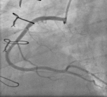
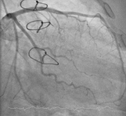
The operator first measured right heart pressures; these were found to be normal with a right atrial pressure of 3 mmHg and preserved cardiac output. The right coronary artery was engaged with a right Judkins 4.0 catheter and appeared angiographically normal (Figure 32-1 and Video 32-1). Using a left Judkins 4.0 catheter, the operator engaged and imaged the left coronary artery; this also appeared angiographically normal (Figure 32-2). At this point, the patient remained symptom-free. Endomyocardial biopsy was performed from the right femoral vein using a 7 French, 98 cm, 40-degree curved sheath positioned into the right ventricle. Three samples were obtained, and while the operator was attempting a fourth sample, the patient developed hypotension and sinus bradycardia. Marked inferior ST-segment elevation appeared on the monitor screen.
The operator engaged a 6 French right Judkins 4.0 guide catheter into the right coronary artery. Angiography demonstrated occlusion of the right coronary artery in the midportion of the vessel (Figure 32-3 and Video 32-2). Another angiogram performed a few minutes later clearly showed air bubbles within the coronary (Figure 32-4
Stay updated, free articles. Join our Telegram channel

Full access? Get Clinical Tree


