CASE 29 Acute Vessel Closure During Coronary Intervention
Cardiac catheterization
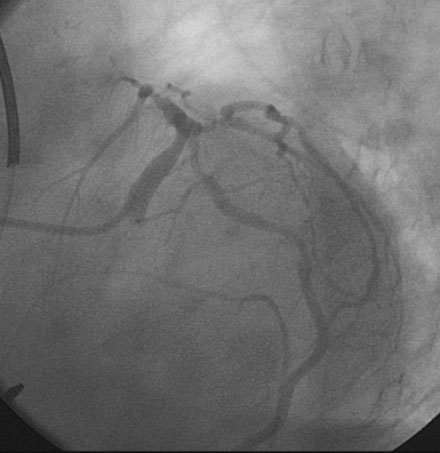
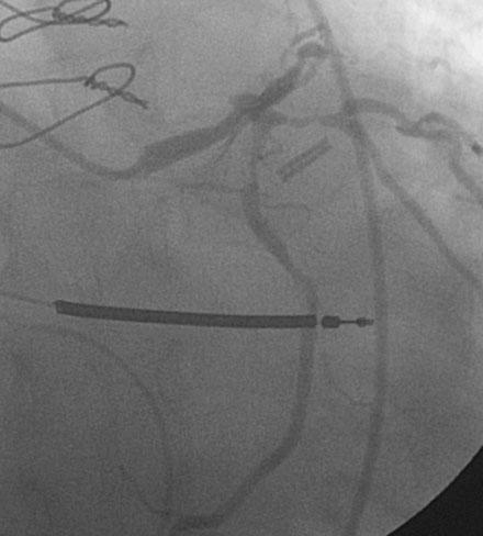
Representative angiograms of her left coronary artery are shown in Figures 29-1, 29-2 and Videos 29-1, 29-2. A complex lesion was present in the proximal portion of the ramus intermedius at a bifurcation. The proximal to midsegment of the circumflex was also severely diseased. Importantly, this artery branched from the left main with marked angulation, clearly seen in the left anterior oblique view with caudal angulation (see Figure 29-1).
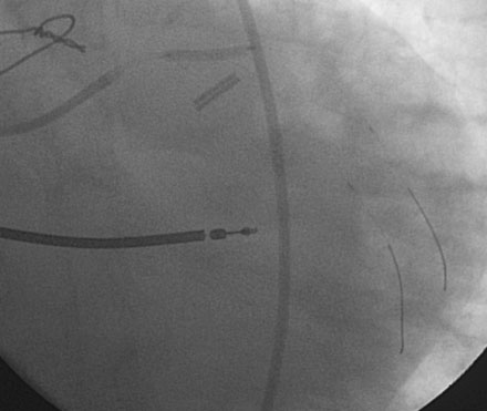
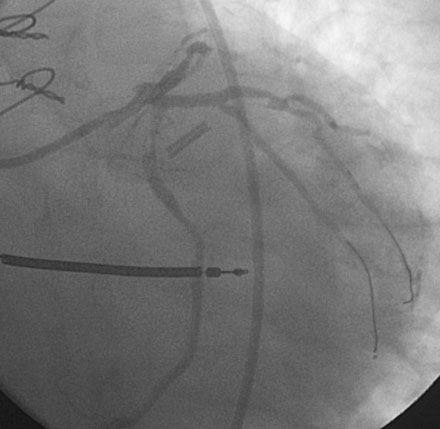
Procedural anticoagulation was accomplished with an intravenous heparin bolus to achieve an activated clotting time greater than 300 seconds and a supportive guide catheter was chosen (6 French JCL 4.5). The operator began with the ramus intermedius (Figure 29-3). This lesion was successfully treated with balloon angioplasty (2.5 mm diameter by 15 mm long compliant balloon) followed by placement of an everolimus-eluting stent (2.75 mm diameter by 18 mm long) (Figure 29-4 and Video 29-3).
Stay updated, free articles. Join our Telegram channel

Full access? Get Clinical Tree


