Chapter 25
Questions
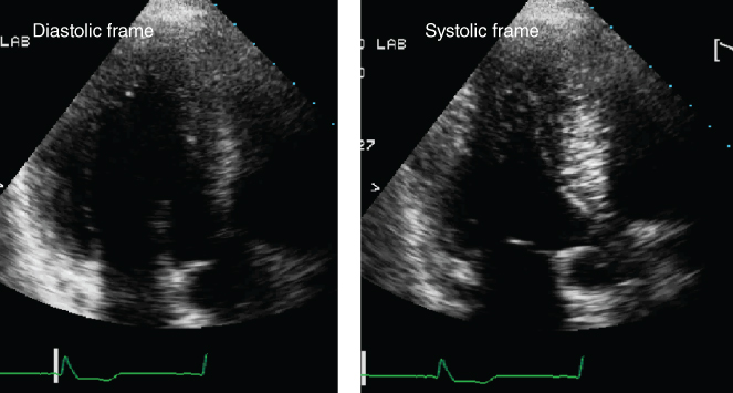
- 481. This still-frame image of a four-chamber view shows:
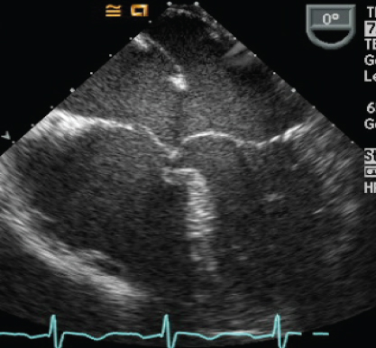
- A. Secundum atrial septal defect
- B. Primum atrial septal defect
- C. Sinus venosus atrial septal defect
- D. None of the above
- 482. The still-frame image of an apical five-chamber view shows:
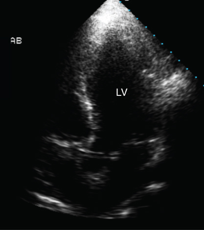
- A. Artifact
- B. Anomalous coronary artery
- C. Coronary sinus
- D. Biventricular pacer lead
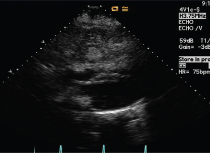
- 483. This parasternal short-axis view shows:
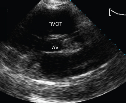
- A. Normal aorta and pulmonary artery
- B. Imaging artifact
- C. Anomalous coronary artery
- D. Artifact from pulmonary prosthesis
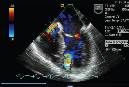
- 484. This is a 51-year-old Armenian male admitted with complaints of chest pain. He underwent an echocardiogram. The still frame of 2D and color images show:
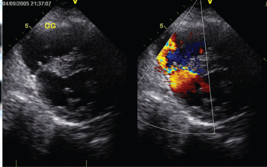
- A. ASD
- B. An inferior septal VSD
- C. Muscular VSD
- D. None of the above
- 485. The continuous wave Doppler shows:
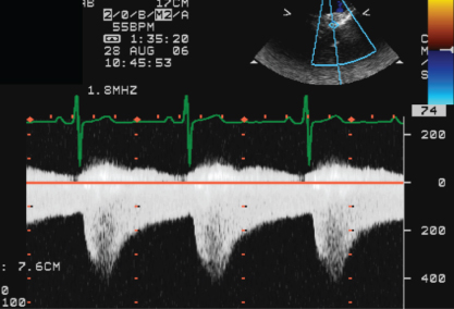
- A. A PDA
- B. Coarctation of the aorta
- C. Coronary fistula
- D. None of the above
- 486. This is a still-frame image of a subcostal view. The image shows:
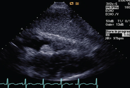
- A. Myxoma
- B. Lipoma
- C. Lipomatous hypertrophy of the interatrial septum
- D. Thrombus attached to the interatrial septum
- 487. This is a still-frame of a four-chamber view. The color flow shows:
- A. Muscular VSD
- B. Apical cannula flow of an LVAD
- C. Psuedoaneurysm
- D. None of the above
- 488. The color flow obtained from parasternal short-axis and suprasternal views show:
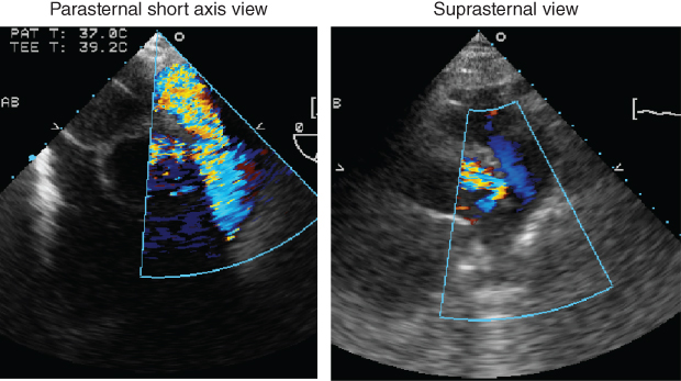
- A. Pulmonary regurgitation
- B. PDA
- C. Coronary fistula
- D. Flow in the coronary artery
- 489. This image was obtained from the subcostal view. This is an image from a 41-year-old male with complaints of diarrhea, flushing, and weight loss. The image shows:
- A. Normal heart and liver
- B. Carcinoid masses in the liver
- C. Liver cysts
- D. None of the above
- 490. This is a still-frame image of an apical long axis image. The image shows:
- A. Normal appearance of the heart
- B. Hypertrophy of the septum
- C. Apical hypertophic cardiomyopathy
- D. Apical thrombus
- 491. The still-frame image of a parasternal short-axis and parasternal long-axis view is shown. This is a 40-year-old male with a history of Marfan syndrome. What surgical procedure did this patient undergo:

- A. Coronary artery bypass
- B. Bentall
- C. Ascending aortic graft
- D. None of the above
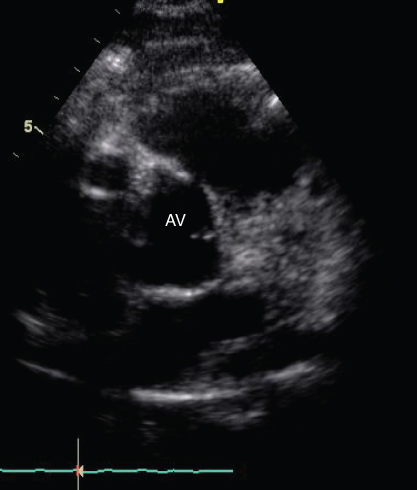
- 492. A 22-year-old male had an echocardiogram as part of routine survellience. The short-axis image shows:

Stay updated, free articles. Join our Telegram channel
- A. Secundum atrial septal defect

Full access? Get Clinical Tree


