Chapter 24
Questions
- 461. A two-chamber view (systolic frame) of the patient is shown in question 460. The arrow points to a defect in:
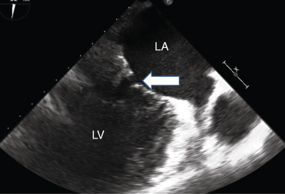
- A. P1 scallop
- B. P2, A2 scallops
- C. P3 scallops
- D. A3 scallop
- 462. The TEE image of the patient in 461 shows:
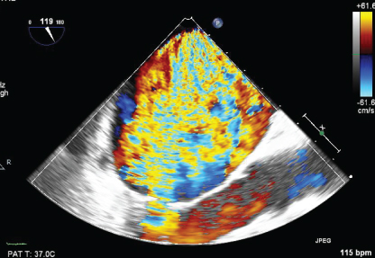
- A. Severe MR
- B. 2+ MR
- C. Severe TR
- D. None of the above
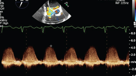
- 463. This pulse Doppler signal from a TEE image from the patient in question 461 is suggestive of:
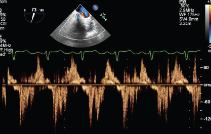
- A. Normal pattern of pulmonary vein pattern
- B. Systolic flow reversal in the pulmonary vein suggestive of severe MR
- C. Systolic flow reversal in the SVC suggestive of severe TR
- D. None of the above
- 464. This is a TEE image from the midesophageal position from patient in question 461. The arrow points to:
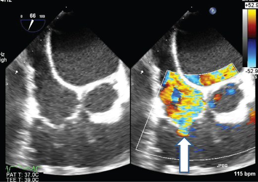
- A. Mild TR
- B. Moderate TR
- C. Severe TR
- D. None of the above
- 465. The CW pattern from the same patient is suggestive of: RA pressure was 20 mm hg
- A. Severe pulmonary hypertension
- B. Moderate pulmonary hypertension
- C. Mild pulmonary hypertension
- D. Cannot be determined
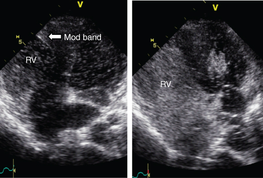
- 466. This is a TEE image of a four-chamber view from a 32-year-old male patient. What is the abnormality noted.
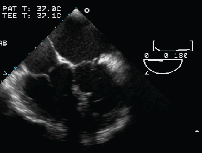
- A. D-TGA
- B. L-TGA
- C. Dextrocardia
- D. Normal heart
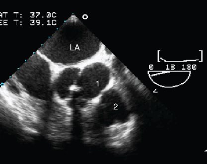
- 467. The numbers 1 and 2 depict which structures?
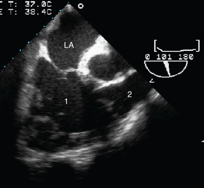
- A. Morphological left ventricle and aorta
- B. Morphological left ventricle and pulmonary artery
- C. Morphological right ventricle and pulmonary artery
- D. Morphological right ventricle and aorta
- 468. The numbers 1 and 2 denote:
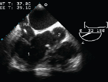
- A. Morphological left ventricle and aorta
- B. Morphological left ventricle and pulmonary artery
- C. Morphological right ventricle and pulmonary artery
- D. Morphological right ventricle and aorta
- 469. The numbers 1 and 2 denote which structures:
- A. Aortic valve, pulmonary valve
- B. Short axis of mitral, tricuspid valves
- C. Pulmonary valve, aortic valve
- D. None of the above
- 470. The diastolic and systolic frame of the pulmonary valve flow is suggestive of:
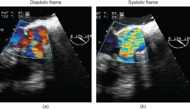
- A. Mild PR only
- B. Mild PR, moderate-to-severe PS
- C. Normal flow pattern
- D. Cannot be determined
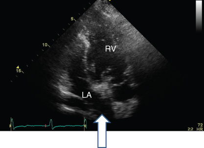
- 471. This is a parasternal long-axis view from a 28-year-old female with c/o shortness of breath. She gives a history of “heart surgery” as a child. The image is suggestive of:
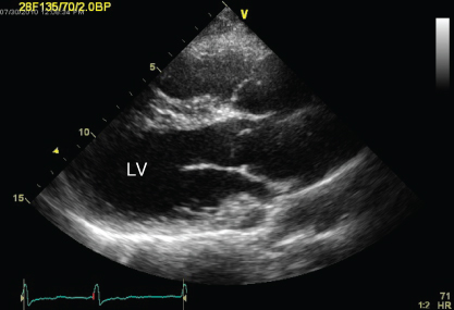
- A. Normal chamber orientation
- B. D-transposition
- C. L-TGA
- D. Cannot determine
- 472. This is a still frame of an apical view. The arrow points to:
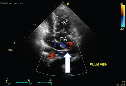
- A. pulmonary vein
- B. SVC
- C. IVC
- D. Pulmonary vein baffle into RA
- 473. This is a still frame of an apical view. The arrow depicts:
- A. IVC
- B. Pulmonary veins
- C. Pulmonary artery
- D. Systemic venous baffle into the LA
- 474. This is an echo image from a 23-year-old female with c/o shortness of breath. She has a h/o D-TGA s/p Mustard procedure. Saline contrast was injected from the arm. The image is suggestive of:

Stay updated, free articles. Join our Telegram channel
- A. P1 scallop

Full access? Get Clinical Tree


