Chapter 22
Questions
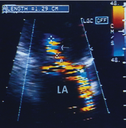
- 421. This TEE image is diagnostic of:
- A. Rheumatic mitral stenosis
- B. Mitral regurgitation
- C. Prosthetic valve stenosis
- D. Calcific mitral stenosis
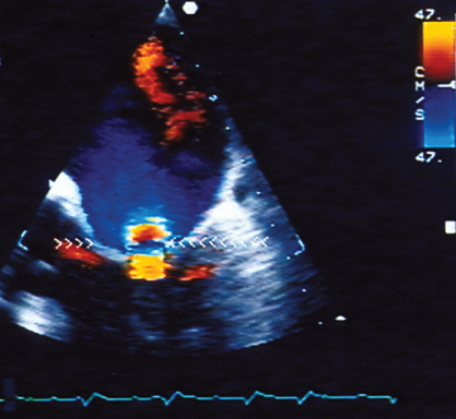
- 422. This transthoracic image is suggestive of:
- A. Mitral stenosis
- B. Mild mitral regurgitation
- C. Severe mitral regurgitation due to flail posterior mitral leaflet
- D. Severe mitral regurgitation due to dilated mitral annulus
- 423. The pulmonary vein flow shown here is suggestive of:
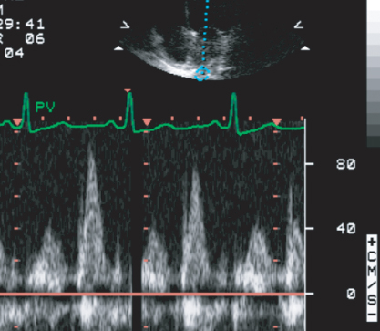
- A. Severe MR
- B. Severe MS
- C. Normal left atrial pressure
- D. High left atrial pressure
- 424. The mitral flow profile and mitral annular velocity in this patient are consistent with:
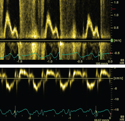
- A. Symptomatic severe MR due to flail mitral valve in a 24-year-old with normal LV size and function
- B. Class III symptoms in a patient with dilated LV and EF of 30%
- C. Normal LV function with mild MR and class I symptoms
- D. Acute severe AR with LVEDP of 55 mmHg
- 425. The parasternal short axis view shown here is consistent with:
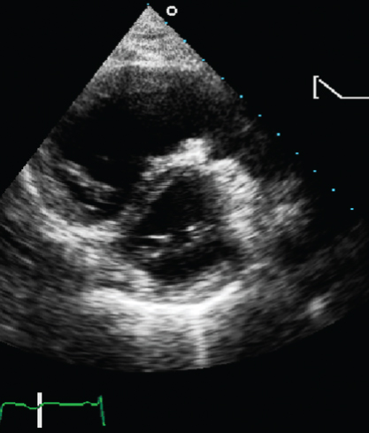
- A. Pulmonary hypertension
- B. Flail mitral valve
- C. Dilated cardiomyopathy
- D. None of the above
- B. Flail mitral valve
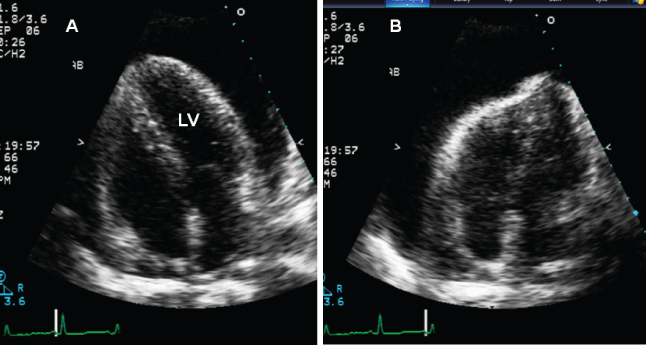
- 426. This patient is likely to have:

- A. Severe LV dysfunction with low cardiac output state
- B. Aortic regurgitation
- C. HOCM
- D. None of the above.
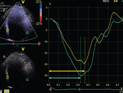
- 427. This patient has:
- A. LV systolic dyssynchrony
- B. LV diastolic dyssynchrony
- C. Good LV synchrony
- D. None of the above
- 428. The signals shown here are annular:
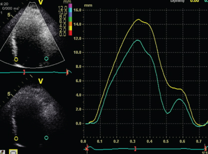
- A. Velocity
- B. Displacement
- C. Strain
- D. Strain rate
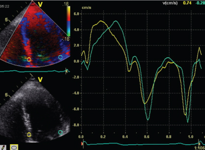
- 429. The signals from septum and LV lateral wall are those of:
- A. LV strain
- B. Strain rate
- C. Velocity
- D. None of the above
- 430. The arrows point to:
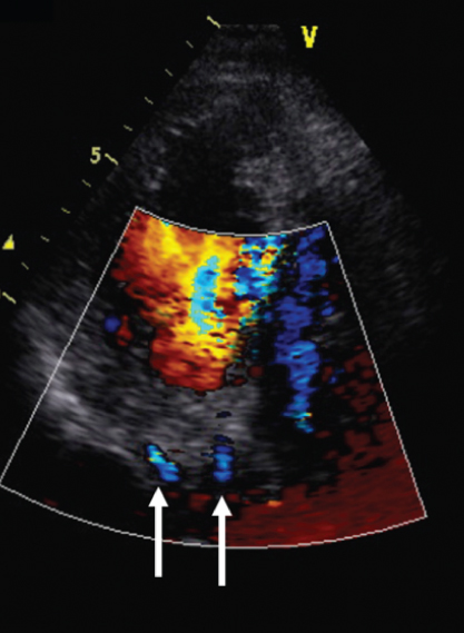
- A. Coronary sinus branches
- B. Coronary artery branches
- C. Artifacts produced by tissue motion
- D. None of the above
- 431. Data shown here permit computation of:
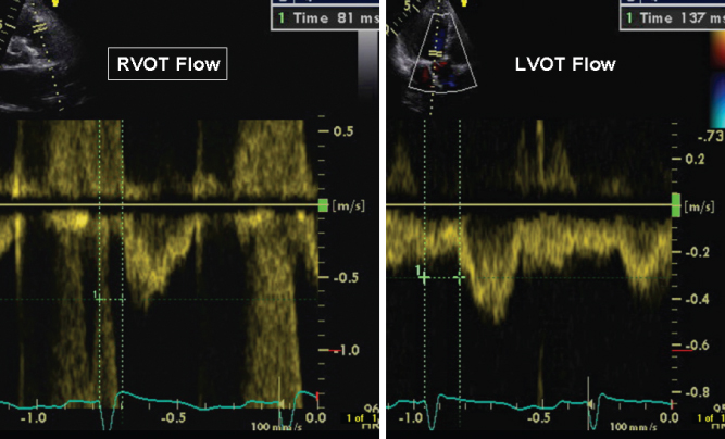
- A. LV intraventricular dyssynchrony
- B. Interventricular dyssynchrony
- C. Atrioventricular dyssynchrony
- D. None of the above
- B. Interventricular dyssynchrony
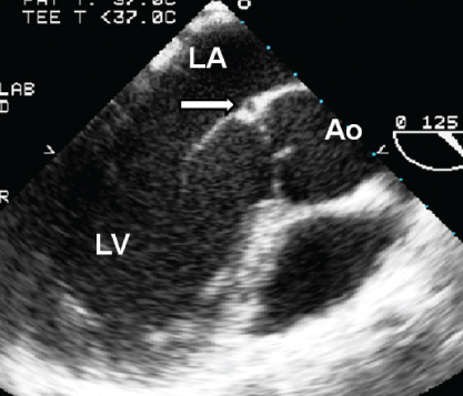
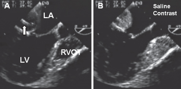
- 432. In this TEE image, downward pointing arrow refers to:
- A. Aortic valve
- B. Vegetation on the aortic valve
- C. Aortic subvalvular membrane
- D. Aortic dissection
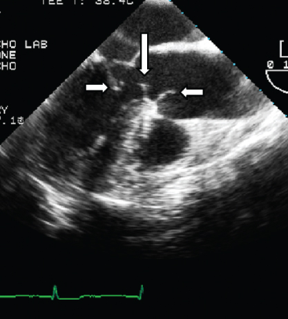
- 433. The arrow on this TEE image points to:
- A. Coronary artery
- B. Aortic valve ring abscess
- C. Artifact
- D. Coronary sinus
- B. Aortic valve ring abscess
- 434. This patient has:
- A. Dilated coronary sinus and dextrocardia
- B. Dilated coronary sinus and levocardia
- C. Cor triatriatum
- D. Aneurysm of circumflex coronary artery
- 435. The arrow points to (DTA = descending thoracic aorta):
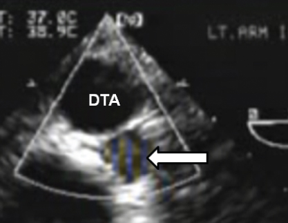
- A. Aortic aneurysm
- B. Inferior vena cava
- C. Dilated azygos vein
- D. Mirror image artifact
- 436. The image shown is indicative of:

Stay updated, free articles. Join our Telegram channel
- A. Rheumatic mitral stenosis

Full access? Get Clinical Tree


