18: Unsuccessful Coronary Intervention

CASE 18 Unsuccessful Coronary Intervention


Case presentation


A 77-year-old woman presented to the hospital emergency department with chest pain at rest. Fourteen years earlier, she underwent coronary bypass surgery consisting of a left internal mammary graft to the left anterior descending artery and saphenous vein grafts to both the right coronary artery and the first obtuse marginal artery. She did well until 1 year earlier when she developed substernal chest pain occurring with exertion and relieved by rest. This symptom progressed and led to diminished exercise tolerance. Her physician evaluated her progressive anginal symptoms by pharmacologic stress perfusion scintigraphy, which confirmed ischemia in the inferior and lateral walls. Based on these results, her physician scheduled her for an outpatient elective cardiac catheterization. However, several prolonged episodes of rest chest pain resolving only after nitroglycerin prompted this hospital visit.


Upon arrival, her chest pain had already resolved and she did not report any other symptoms. In addition to the prior coronary bypass surgery, review of her past medical history was notable for the presence of a left bundle branch block on electrocardiogram, Factor V Leiden with several occurrences of deep vein thrombosis, hypercholesterolemia, and prior tobacco abuse. Home medications included warfarin, atenolol, aspirin, and fluvastatin. Physical examination found no abnormalities. Routine laboratory evaluation confirmed normal renal function, a hematocrit of 43%, and an International Normalized Ratio (INR) of 2.4. Initial troponin I was 0.02 ng/mL, subsequently increasing to 0.21 ng/mL. She was admitted to a telemetry unit and treated with unfractionated heparin in addition to aspirin, beta blockers, and nitrates; warfarin was held to allow the INR to return to baseline, at which point she underwent cardiac catheterization.


Cardiac catheterization


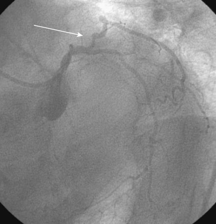
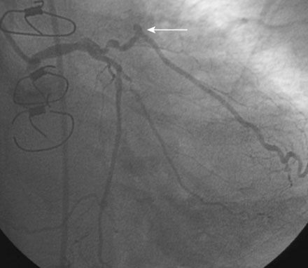
The diagnostic angiograms demonstrated occlusion of the native right coronary and left anterior descending coronary artery with occlusion of both saphenous vein grafts. A widely patent left internal mammary graft supplied a large left anterior descending artery. Severe disease of the proximal segment of a large first obtuse marginal artery as well as severe disease of a smaller-caliber second obtuse marginal artery likely accounted for her clinical presentation (Figures 18-1, 18-2 and Video 18-1). Based on these angiograms, stress test results, and her symptoms of progressive angina, her physician decided to revascularize the first obtuse marginal artery percutaneously.


Jun 11, 2016 | Posted by in CARDIOLOGY | Comments Off on 18: Unsuccessful Coronary Intervention

Full access? Get Clinical Tree

Get Clinical Tree app for offline access