CASE 17 Extensive Coronary Dissection
Cardiac catheterization
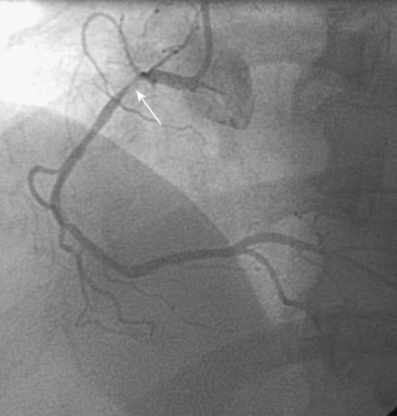
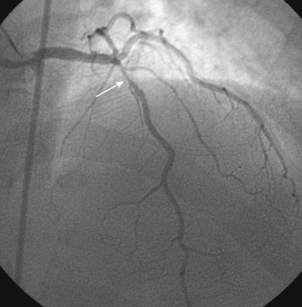
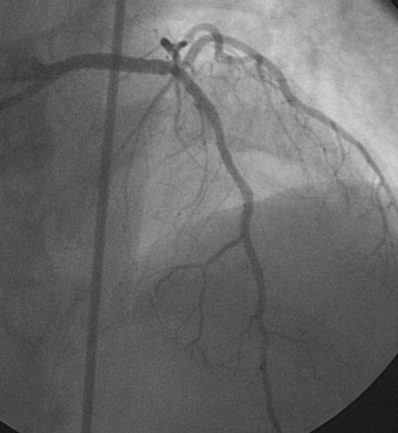
Cardiac catheterization revealed preserved left ventricular systolic function and multivessel coronary disease, with severe discrete stenoses of the proximal right coronary artery and left anterior descending artery and an angiographically normal circumflex artery (Figures 17-1, 17-2 and Videos 17-1, 17-2). The attending physician discussed revascularization options, and the patient chose percutaneous coronary intervention of both the right coronary and left anterior descending coronary arteries with drug-eluting stents instead of bypass surgery. She returned to the cardiac catheterization laboratory the next morning following a dose of 300 mg of clopidogrel taken the night before. The operator began with the left anterior descending artery. This was successfully treated with a 2.5 mm diameter by 18 mm long, drug-eluting stent (zotarolimus), with an excellent angiographic result (Figure 17-3). The operator engaged a 6 French right Judkins 4.0 guide catheter in the right coronary artery and easily crossed the stenosis with a floppy-tipped guidewire and predilated the lesion to 10 atmospheres with a 2.5 mm diameter by 15 mm long compliant balloon. The operator then attempted, but failed to pass, a 2.5 mm diameter by 24 mm long drug-eluting stent (zotarolimus) across the lesion. The stent met significant resistance at the site of a bend in the proximal right coronary artery (see Figure 17-1, arrow), and the attempt caused the guide to inadvertently disengage, resulting in loss of guidewire access to the distal right coronary artery. Angiography following reengagement of the guide catheter showed no obvious dissection but a persistent residual stenosis at the site of the balloon-dilated lesion (Video 17-3).