16: Acute Versus Chronic Thromboembolic Disease—Beware!


Figure 16.1

Transthoracic echocardiogram showing signs of right ventricular strain


This was followed by the acquisition of a CT angiogram of the chest. This revealed bilateral pulmonary emboli affecting the proximal lobar vessels of the right upper and bilateral lower lobes, extending into the segmental branches. Partially occluding thrombi are seen within the segmental left upper lobe branches and a large thrombus is visible in the left lower lobe pulmonary artery extending into the segmental branches. There was a small infiltrate versus atelectasis in the anterior left lung base, and a small amount of anterior pericardial fluid (Fig. 16.2).

../images/1_1_En_16_Chapter/1_1_En_16_Fig2_HTML.png

Figure 16.2

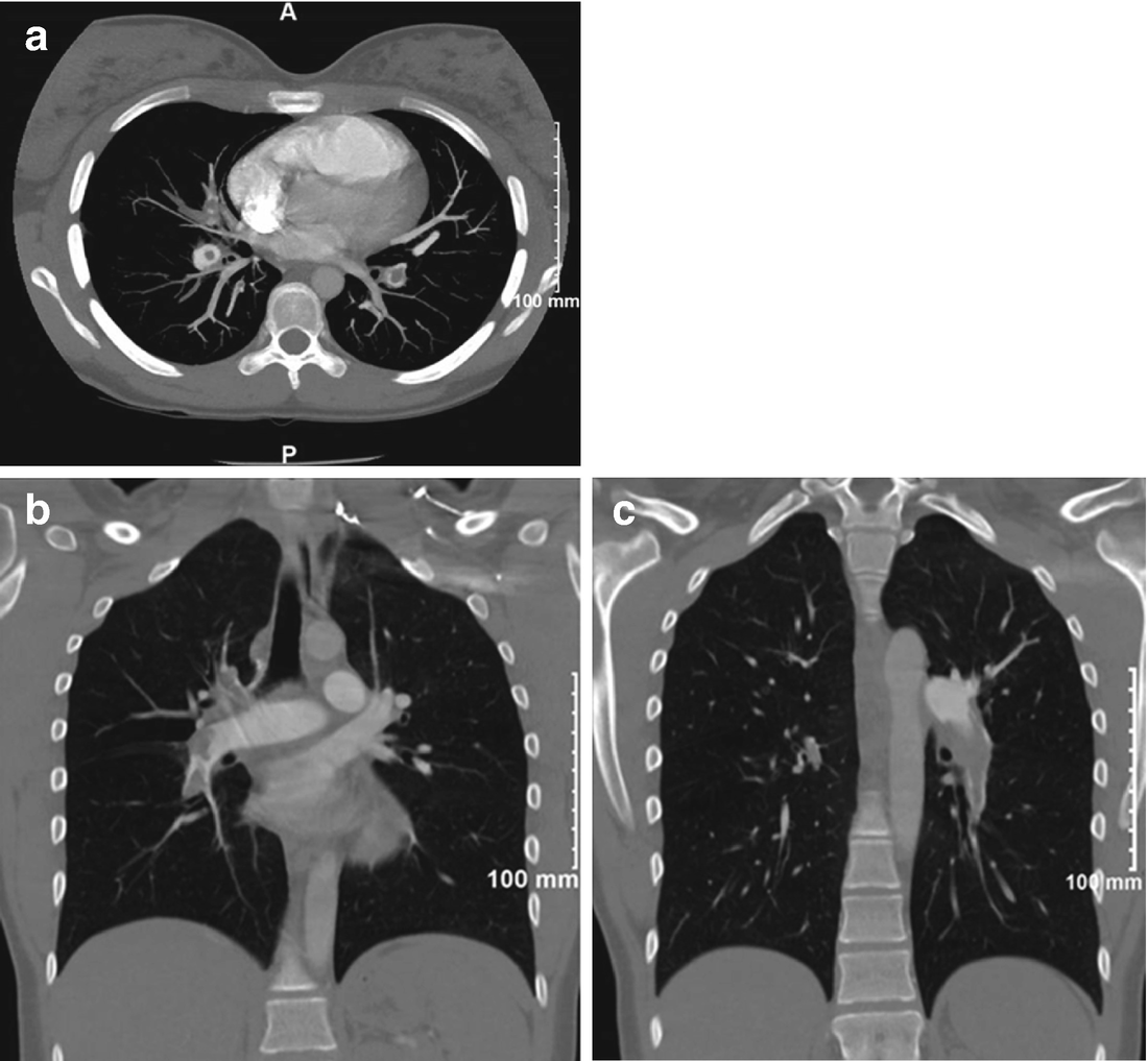
Axial (a) and coronal views (b, c) of the CT angiogram demonstrating extensive proximal vessel intraluminal defects in both lungs consistent with acute pulmonary emboli. There is modest central PA enlargement and RV enlargement, though no significant RV hypertrophy


A duplex venous ultrasound of both lower extremities disclosed the deep veins to be widely patent with normal flow and no evidence of thrombus.


She was admitted to the Intensive Care Unit, after initiating intravenous heparin, with the intention of proceeding with lytic therapy utilizing tissue plasminogen activator (TPA). The clinical history related was consistent with a possible pulmonary embolic event as long as 6 weeks prior; however her presentation was prompted by worsening symptoms that were hemodynamically significant.


She was transferred to a tertiary hospital as the patient and parents requested additional consults prior to moving forward with thrombolysis given lack of consensus in the treatment plan. Hematology and Pulmonary Hypertension/Cardiology consults were obtained and achieved consensus was to withhold lytic therapy for concern that she had acute on chronic pulmonary emboli.


A lung ventilation-perfusion scan was recommended. (Hospital Day # 3) This showed extensive modest to large sized peripherally located wedge shaped perfusion defects bilaterally throughout the lungs, consistent with findings of pulmonary emboli by CT (Fig. 16.3).

../images/1_1_En_16_Chapter/1_1_En_16_Fig3_HTML.png

Figure 16.3

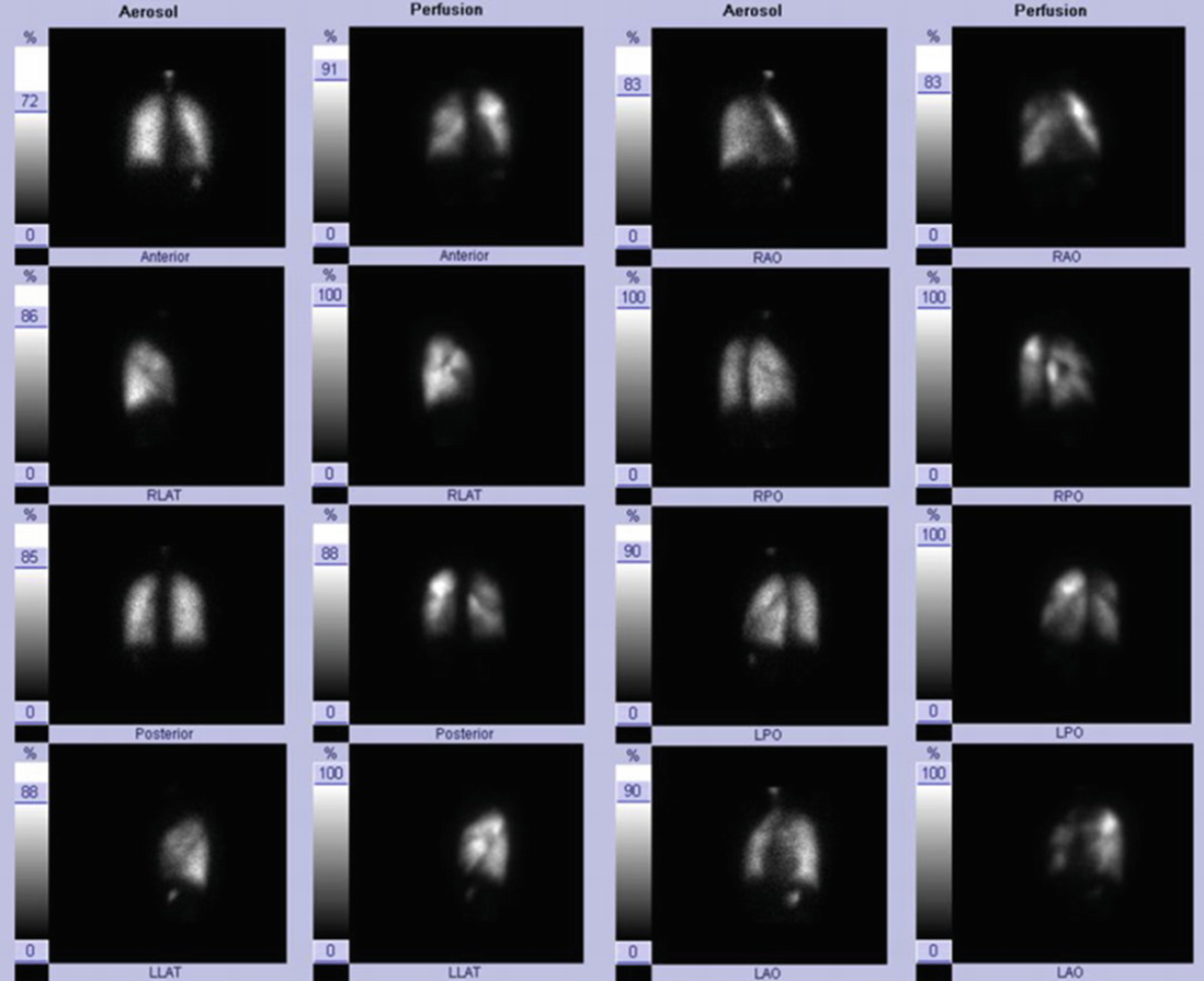
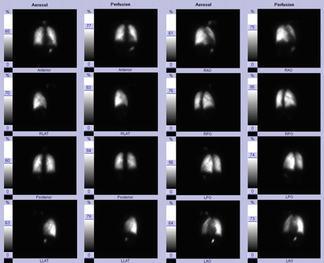
Several unmatched, large perfusion defects, particularly apparent throughout the right lung and medial left lower lobe (posterior view)


On hospital day 6, a right heart catheterization was performed: BP 99/66 mmHg, mean arterial pressure 79 mmHg, HR 77 bpm, pulse oximetry on room air 99%. RA mean 6 mmHg, RV 40/6 mmHg, PA pressure 40/17 (25 mean) mmHg, PCWP 6 mmHg; PA saturation 67.3%; cardiac output 4.5 L/min with a cardiac index 2.43 L/min/m2; transpulmonary gradient was 19 mmHg.


Concurrent with her catheterization, pulmonary angiography was obtained. This demonstrated pronounced filling defects in the left lower pulmonary arteries and smaller filling defects in the segmental and subsegmental levels; there were filling defects in the right upper, middle, and lower lobe pulmonary arteries at the lobar and segmental levels (Fig. 16.4).

../images/1_1_En_16_Chapter/1_1_En_16_Fig4_HTML.jpg

Figure 16.4

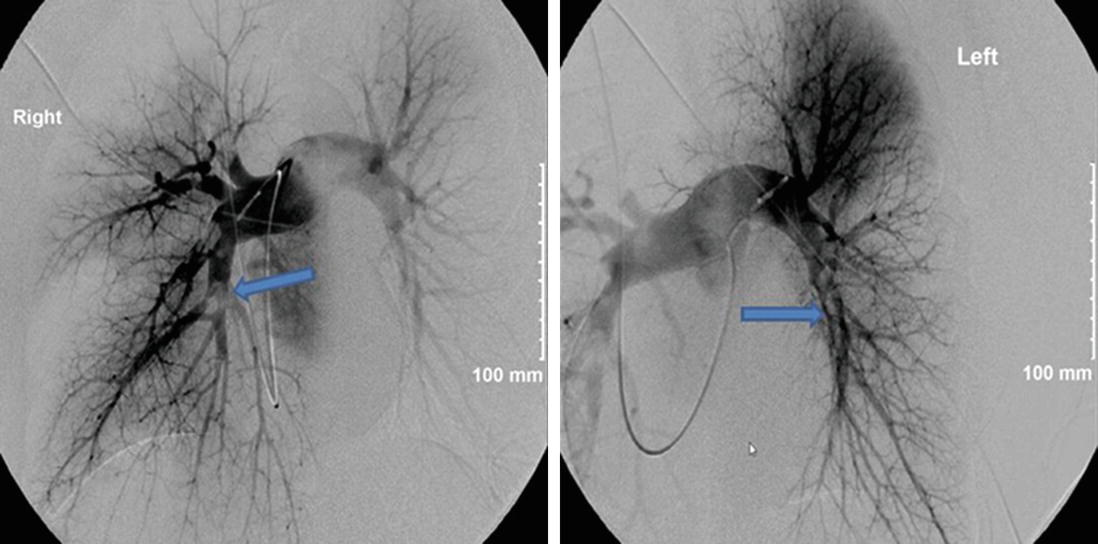
Intraluminal filling defects apparent (arrows) in both descending pulmonary arteries, as well as in the segmental vessels of the right upper lobe, right lower lobe and lingula


By hospital day 12, she was discharged in stable condition. She was at a therapeutic level on warfarin therapy (patient refused Eliquis). She was hemodynamically stable and ambulatory. Thrombolytic therapy was never given, as the consensus of opinions was that she had acute pulmonary emboli on top of significant chronic thrombi. The oral contraceptive (OCP) drugs were discontinued on admission.


Six weeks after hospital discharge the patient was working full-time in a restaurant in her hometown, was increasing her activities and doing well. She was stable on 8 mg of warfarin daily. By eleven weeks after hospital discharge, the patient was feeling fine with no complaints. She was participating in a yoga class and sailing. She also underwent a HAST study for a planned vacation trip to San Diego and did not require oxygen for the flight. She denied dyspnea with her activities, though still had not regained her usual physical activity level yet. Her physical examination was unremarkable. Hypercoagulable lab studies all came back negative. A limited echocardiogram showed normal RV size, with mildly decreased systolic function (40%), and a PA systolic pressure estimated at 23 mmHg, Chest radiograph findings were “normal” and a lung ventilation-perfusion scan documented resolution of the previously observed perfusion defects (Fig. 16.5).

../images/1_1_En_16_Chapter/1_1_En_16_Fig5_HTML.png

Only gold members can continue reading. Log In or Register to continue

Stay updated, free articles. Join our Telegram channel

Oct 30, 2020 | Posted by in Uncategorized | Comments Off on 16: Acute Versus Chronic Thromboembolic Disease—Beware!

Full access? Get Clinical Tree

Get Clinical Tree app for offline access