Chapter 15
Questions
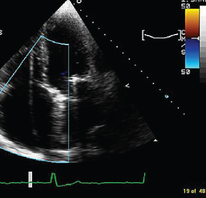
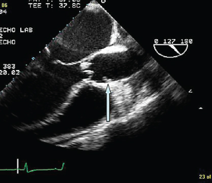
- 281. This image shows a vegetation on the:
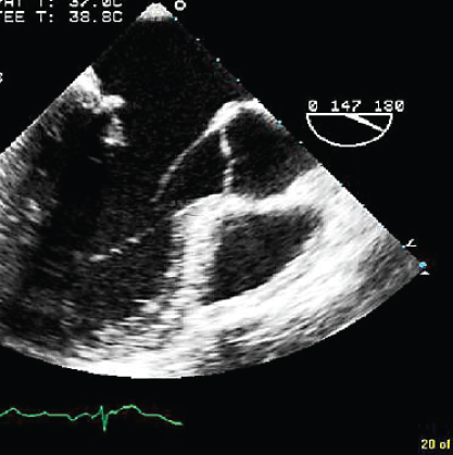
- A. Aortic valve
- B. P2 scallop of mitral valve
- C. P1 scallop of mitral valve
- D. A2 scallop of mitral valve
- 282. The hemodynamics in this patient potentially could be improved by:
- A. Shortening the PR interval
- B. Afterload reduction
- C. Positive inotropes
- D. All of the above
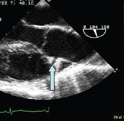
- 283. The trans-esophageal echocardiogram (TEE) image shown here is indicative of:
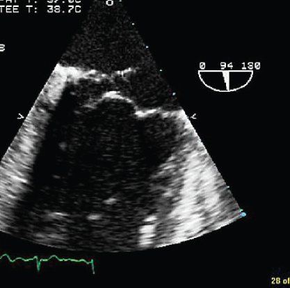
- A. Flail posterior leaflet P3 segment
- B. Flail posterior leaflet P1 segment
- C. Flail anterior leaflet
- D. Large mitral valve vegetation
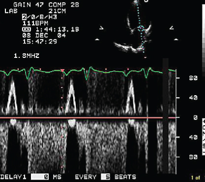
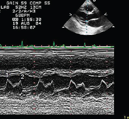
- 284. The pulse wave Doppler in the right upper pulmonary vein is indicative of:
- A. Abnormal left ventricular (LV) relaxation
- B. High left atrial (LA) pressure
- C. Mitral stenosis
- D. Severe mitral regurgitation (MR)
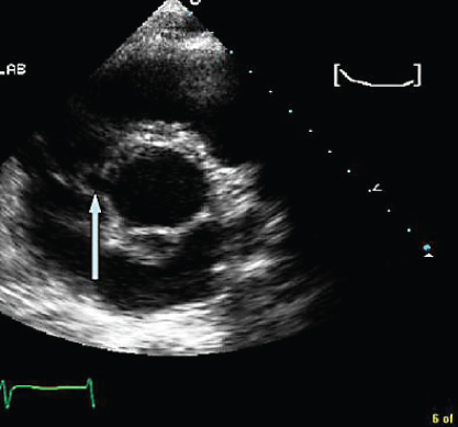
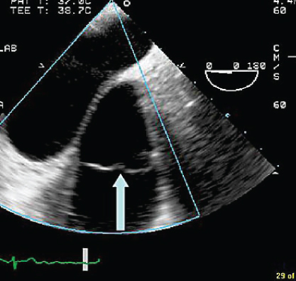
- 285. This apical four-chamber view shows:
- A. A pacemaker lead in the right ventricle (RV)
- B. A pacemaker lead in the coronary sinus
- C. Epicardial RV lead
- D. Artifact in the RV
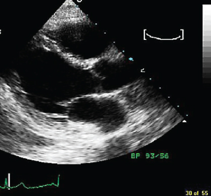
- 286. The mitral valve opening pattern in this patient is suggestive of:
- A. Mitral stenosis
- B. High left ventricular end diastolic pressure (LVEDP)
- C. Atrial fibrillation
- D. Normal pattern
- 287. The part of the anatomy and measurement indicated by the line is:
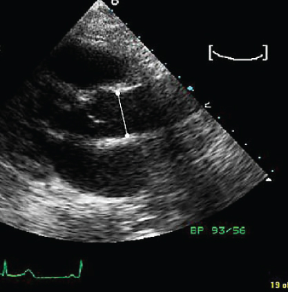
- A. The sino-tubular junction
- B. Sinus diameter
- C. Sinus height
- D. Aortic annular diameter
- B. Sinus diameter
- 288. The blood supply to the ventricular septum shown here is:
- A. Left anterior descending (LAD)
- B. Posterior descending artery
- C. Both
- D. Neither
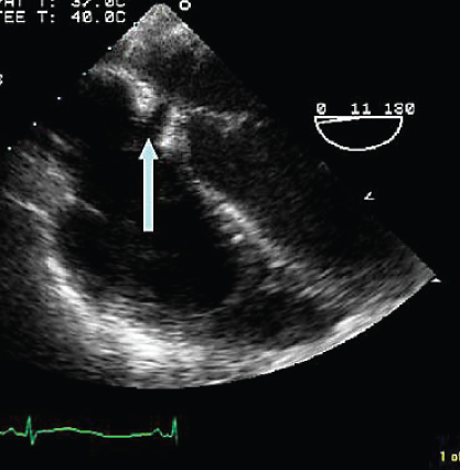
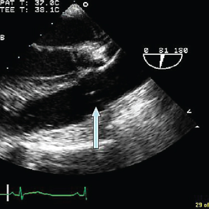
- 289. The structure indicated by the arrow in the ascending aorta is likely to be:
- A. Vegetative aortitis
- B. Flap of aortic dissection
- C. Intraaortic atherosclerotic debris
- D. Supravalvular aortic stenosis
- B. Flap of aortic dissection
- 290. The structure indicated by the arrow is likely to be:
- A. Aortic dissection
- B. Aortic transaction
- C. Right coronary artery
- D. Left coronary artery
- 291. The arrow in this short axis view transthoracic echocardiogram (TTE) image at the level of the ascending aorta is:
- A. Artifact
- B. Tissue plane and aorta and RV outflow tract
- C. Aortic dissection
- D. Right coronary artery
- B. Tissue plane and aorta and RV outflow tract
- 292. The structure shown by the arrow is:
- A. Coronary sinus
- B. Atrial septal defect (ASD)
- C. Superior vena cava
- D. Inferior vena cava
- 293. The valve indicated by the arrow is:
- A. Pulmonary valve
- B. Aortic valve
- C. Tricuspid valve
- D. Mirror image artifact of the aortic valve
- B. Aortic valve
- 294. This view is obtained from the upper esophagus. The structure indicated by the arrow is:
- A. Aortic valve
- B. Pulmonary valve
- C. Tricuspid valve
- D. Artifact
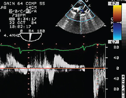
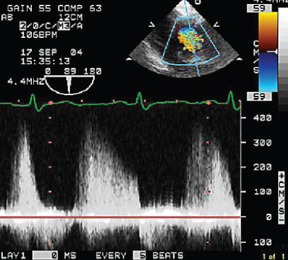
- 295. The pulmonary regurgitation signal shown here is indicative of (assuming right atrial pressure of 15 mmHg):

Stay updated, free articles. Join our Telegram channel
- A. Aortic valve

Full access? Get Clinical Tree


