Chapter 14
Questions
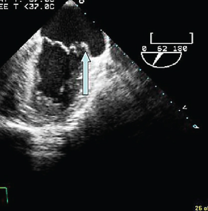
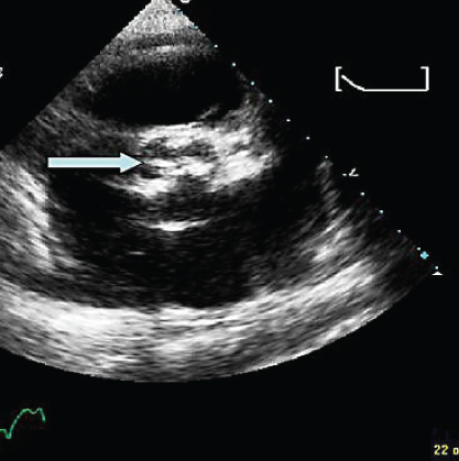
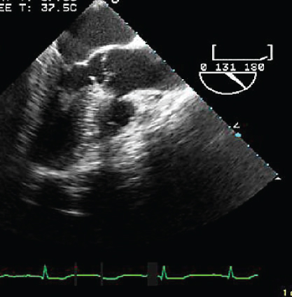
- 261. The arrow here points to:
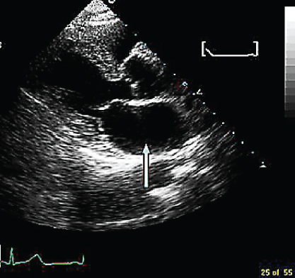
- A. Left atrium
- B. Right pulmonary artery
- C. Posterior pericardial effusion
- D. Left pleural effusion
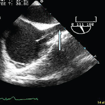
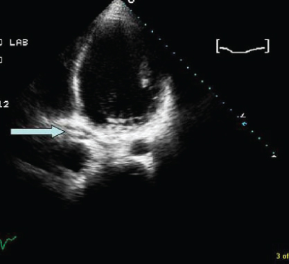
- 262. The structure denoted by the arrow is:
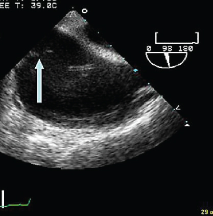
- A. Vegetation
- B. Eustachian valve
- C. Edge of atrial septal defect (ASD)
- D. Tricuspid valve
- 263. The structure shown by the arrow is:
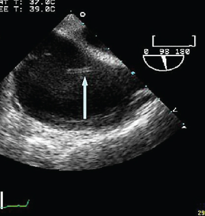
- A. Artifact
- B. Catheter in right atrium
- C. Thrombus
- D. Loose suture material
- B. Catheter in right atrium
- 264. The patient may have all of the following except:
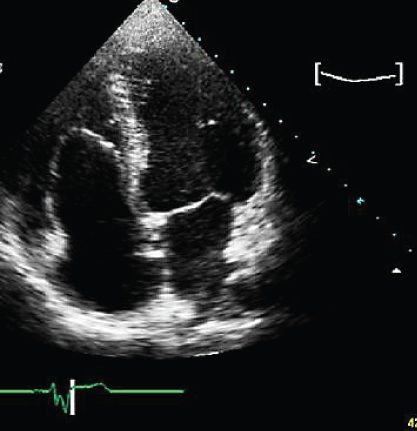
- A. Atrial septal defect
- B. Wolf–Parkinson–White syndrome
- C. Tricuspid regurgitation
- D. Bicuspid aortic valve
- 265. The mitral valve abnormality seen here is:
- A. Perforation, prolapse of P1 scallop of posterior leaflet
- B. Abnormal P3 scallop
- C. Prolapsing P2 scallop
- D. Anterior leaflet prolapse
- B. Abnormal P3 scallop
- 266. The structure denoted here is:
- A. Superior vena cava
- B. Inferior vena cava
- C. Right upper pulmonary vein
- D. Main pulmonary artery
- 267. The numbers 1, 2, and 3 denote the following cusps of the aortic valve:
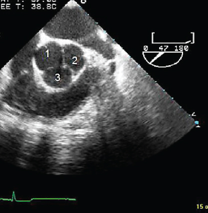
- A. Non, left, right coronary cusps
- B. Left, right, non-coronary cusps
- C. Right, left, non-coronary cusps
- D. Non-coronary, right, left cusps
- B. Left, right, non-coronary cusps
- 268. Structure no. 4 denotes:
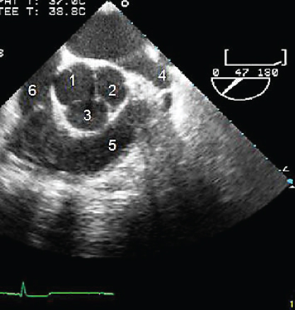
- A. Left atrial appendage
- B. Right atrial appendage
- C. Left upper pulmonary vein
- D. Left lower pulmonary vein
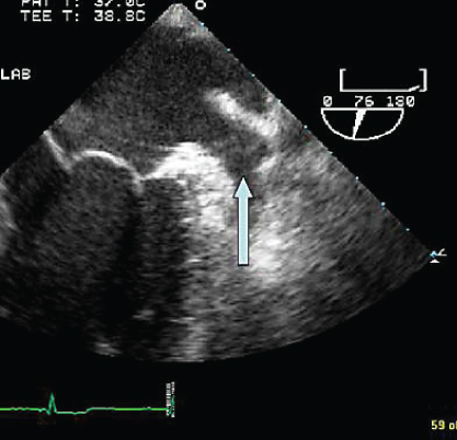
- 269. The structure shown by the arrow is:
- A. Calcified native aortic valve
- B. Stented bioprosthetic aortic valve
- C. St. Jude bileaflet mechanical aortic valve
- D. Supravalvular aortic stenosis as part of William’s syndrome
- B. Stented bioprosthetic aortic valve
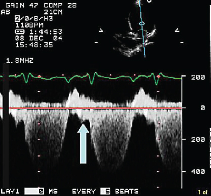
- 270. The M-mode echocardiogram is suggestive of:
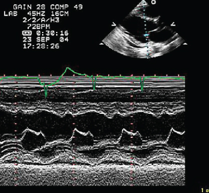
- A. Normal mitral valve motion
- B. Mitral stenosis
- C. Severe aortic regurgitation
- D. High left atrial pressure
- 271. The image shown here is suggestive of:
- A. Mitral annuloplasty
- B. Catheter in the coronary artery
- C. Biventricular pacemaker or ICD
- D. An artifact
- B. Catheter in the coronary artery
- 272. The structure denoted by the arrow is:
- A. Left atrial appendage
- B. Left lower pulmonary vein
- C. Left upper pulmonary vein
- D. Right lower pulmonary vein
- 273. The patient shown here has:
- A. Valvular aortic stenosis
- B. Subvalvular aortic stenosis
- C. Endocarditis
- D. Hypertrophic obstructive cardiomyopathy
- B. Subvalvular aortic stenosis
- 274. The arrow is indicative of:
- A. Diastolic mitral regurgitation
- B. An artifact
- C. Pulmonary vein D wave picked up by the continuous wave cursor
- D. Mitral annular motion superimposed on the mitral flow
- 275. This patient is likely to have:


Stay updated, free articles. Join our Telegram channel
- A. Left atrium

Full access? Get Clinical Tree


