Chapter 13
Questions
- 241. This patient is likely to have:

- A. Severe aortic stenosis (AS)
- B. Severe mitral regurgitation (MR)
- C. Severe pulmonary hypertension
- D. Mild AS
- 242. For the patient in question 241 the left ventricular outflow tract (LVOT) diameter was 2 cm and the LVOT velocity by pulse Doppler was 1 m/s. The aortic valve area by the continuity equation would be:
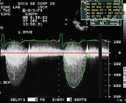
- A. 0.2 cm2
- B. 0.3 cm2
- C. 0.5 cm2
- D. 0.8 cm2
- B. 0.3 cm2
- 243. Image of the aortic arch shown here is indicative of:
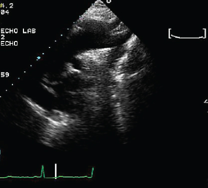
- A. Aneurysm of the aortic arch
- B. Aortic dissection
- C. Severe coarctation of the aorta
- D. Stented aortic coarctation
- 244. This is the continuous wave signal obtained from the pulmonary valve at the mid- to proximal esophageal location. This patient is likely to have:
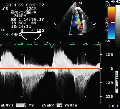
- A. Wide open pulmonary regurgitation (PR)
- B. Mild PR
- C. Severe valvular pulmonary stenosis (PS)
- D. Severe subvalvular PS
- B. Mild PR
- 245. This patient has vegetation on:
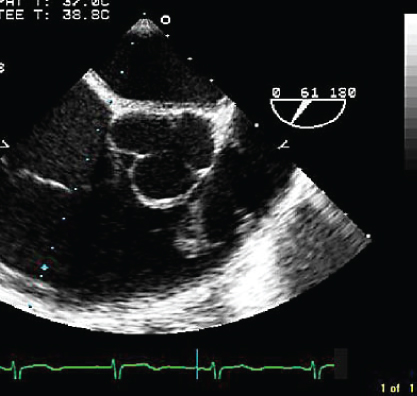
- A. Aortic valve
- B. Pulmonary valve
- C. Tricuspid valve
- D. Pacemaker lead
- 246. The appearance of the left atrial cavity is caused by:

- A. Stasis of blood
- B. Mitral regurgitation
- C. Polycythemia
- D. Hyperdynamic circulation
- 247. The cause of the patient’s mitral valve problem is:
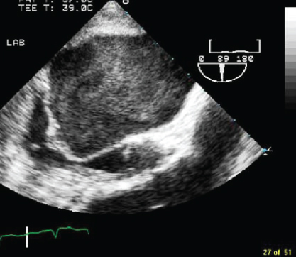
- A. Rheumatic heart disease
- B. Degenerative valve disease
- C. Fen Phen valvulopathy
- D. Ischemic heart disease
- 248. The arrow in this image points to:
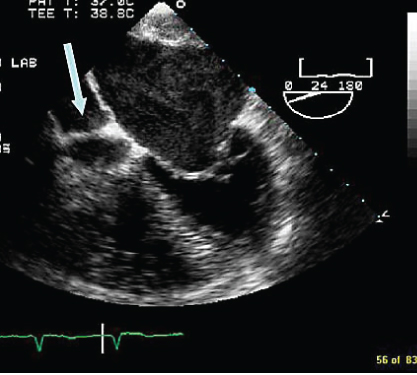
- A. Right atrium (RA)
- B. Coronary sinus
- C. Left atrium (LA)
- D. Right ventricle (RV)
- 249. The arrow in this image points to:
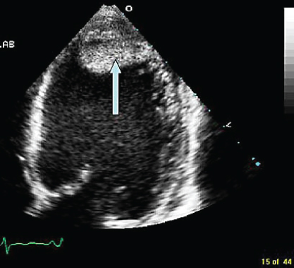
- A. Left ventricular (LV) apical thrombus
- B. RV thrombus
- C. Rib artifact
- D. LA thrombus
- 250. This patient is likely to have:
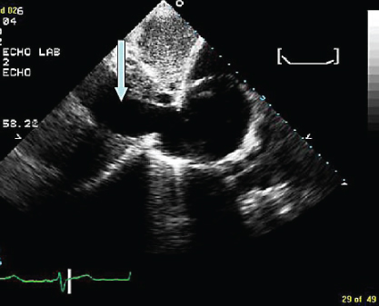
- A. High RA pressure
- B. Pericardial effusion
- C. Aortic dissection
- D. Dilated azygos vein
- 251. The pulmonary vein flow shown here is indicative of:
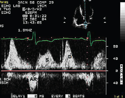
- A. Elevated LA pressure with normal end diastolic pressure (EDP)
- B. Elevated LA pressure with elevated EDP
- C. Abnormal LV relaxation with normal EDP
- D. Elevated LVEDP with normal LA pressure
- 252. The mitral flow pattern shown here is suggestive of:
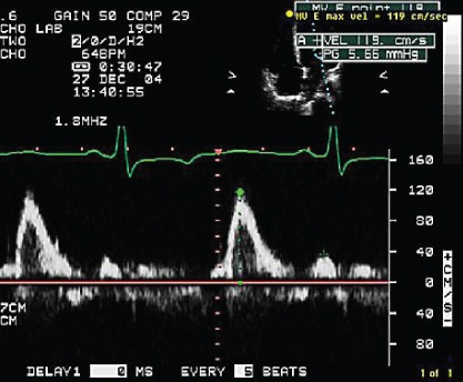
- A. Normal LA pressure
- B. High LA pressure
- C. Atrial mechanical failure
- D. Abnormal LV relaxation with normal LA pressure
- 253. This patient has:
- A. Mitral atresia
- B. Tricuspid atresia
- C. Transposition of great vessels with atrial baffle
- D. Epstein’s anomaly

Stay updated, free articles. Join our Telegram channel
- A. Severe aortic stenosis (AS)

Full access? Get Clinical Tree


