, Marilyn J. Siegel2, Tomasz Miszalski-Jamka3, 4 and Robert Pelberg1
(1)
The Christ Hospital Heart and Vascular Center of Greater Cincinnati, The Lindner Center for Research and Education, Cincinnati, OH, USA
(2)
Mallinckrodt Institute of Radiology, Washington University School of Medicine, St. Louis, Missouri, USA
(3)
Department of Clinical Radiology and Imaging Diagnostics, 4th Military Hospital, Wrocław, Poland
(4)
Center for Diagnosis Prevention and Telemedicine, John Paul II Hospital, Kraków, Poland
Abstract
In a normal biventricular heart, the systemic and pulmonary circulations are in series and each circulation is supported by its respective ventricle. In patients with single ventricular chamber morphology, the two circulations are in parallel and patients only survive by mixing of the systemic and pulmonary venous blood, usually via a septal defect. Systemic venous to pulmonary artery surgical connections have been created to provide venous flow to the pulmonary circulation to allow oxygenation of the blood.
In a normal biventricular heart, the systemic and pulmonary circulations are in series and each circulation is supported by its respective ventricle. In patients with single ventricular chamber morphology, the two circulations are in parallel and patients only survive by mixing of the systemic and pulmonary venous blood, usually via a septal defect. Systemic venous to pulmonary artery surgical connections have been created to provide venous flow to the pulmonary circulation to allow oxygenation of the blood.
25.1 Glenn Shunt
The original (classic) Glenn shunt is an end-to-end anastomosis of the superior vena cava to the right pulmonary artery, which is divided from the main pulmonary artery. All proximal superior vena cava blood flow is directed to the right pulmonary artery (Figs. 25.1 and 25.2). The azygous vein is also ligated. This shunt was used to palliate congenital heart defects with right-sided hypoplasia or atresia such as tricuspid atresia, Epstein anomaly, and pulmonary atresia with intact ventricular septum.

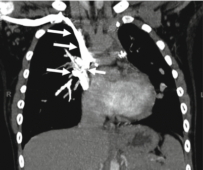
Fig. 25.1
Glenn shunt. This shunt is an end-to-end anastomosis of the right pulmonary artery (RPA), which is divided from the main pulmonary artery to the superior vena cava (SVC), directing all proximal SVC blood flow to the RPA. The SVC is ligated at its entrance to the right atrium (RA). Ao aorta, MPA main pulmonary artery, RAA right atrial appendage, RPA right pulmonary artery
Fig. 25.2
Glenn shunt for tricuspid atresia. This coronal image shows the shunt (white arrows), which extends from the superior vena cava to right pulmonary artery
The modified Glenn shunt, also known as the bidirectional Glenn shunt, consists of an end-to-side anastomosis of the superior vena cava to the right pulmonary artery, which is not divided from the main pulmonary artery (Figs. 25.3 and 25.4). Because the right pulmonary artery maintains continuity with the main pulmonary artery, blood flows from the superior vena cava into both the right and left pulmonary arteries. The complication of both the original and modified Glenn shunts is the development of pulmonary arteriovenous malformations. Currently, the modified Glenn shunt is used as a staging procedure in children with single-ventricle physiology who will later undergo a Fontan procedure.
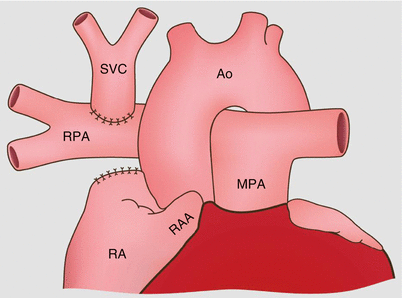
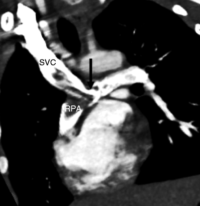
Fig. 25.3
Modified or bidirectional Glenn shunt. This shunt is an end-to-side anastomosis between the superior vena cava (SVC) and the right pulmonary artery. The SVC is divided from the right atrium. The right pulmonary artery is not separated from the main pulmonary artery (MPA) so venous blood flows into both the right and left pulmonary arteries. RAA right atrial appendage, RPA right pulmonary artery, Ao aorta. This procedure is used in patients with single-ventricle physiology who eventually will undergo a Fontan procedure
Fig. 25.4
Bidirectional Glenn shunt. This coronal view shows the end-to-side anastomosis between the superior vena cava (SVC) and the right pulmonary artery (RPA) at the junction with the main pulmonary artery (arrow). Note that contrast-enhanced blood flows to both the right and left pulmonary arteries. The non-opacified blood from the superior vena cava is a flow artifact (not thrombus). Artifact can be differentiated from thrombus by reimaging at 2 min which allows enough time for optimal venous opacification
25.2 Fontan Operation
In 1971, Francis Fontan and Eugene Baudet described a procedure that diverted all systemic venous blood into the pulmonary arteries without the interposition of a ventricle as surgical palliation for tricuspid atresia. The Fontan procedure revolutionized the treatment of complex congenital heart defects and remains the palliative treatment of choice for patients born with one functional ventricle. The primary aim of the Fontan procedure is to establish a circulation in which the systemic venous return enters the pulmonary arteries directly.
Creation of the Fontan circulation is considered in all patients with complex congenital heart disease when a biventricular repair is not possible. These include patients with tricuspid atresia, pulmonary atresia with intact ventricular septum, double-inlet left ventricle, hypoplastic left heart syndrome, double-outlet right ventricle, and complete unbalanced atrioventricular septal defects. Candidates for Fontan procedure should be in sinus rhythm and have adequately developed central pulmonary arteries and good ventricular function. The absence of any one of these criteria is a strong predictor of an early poor outcome.
The classic Fontan operation consists of a valved conduit between the right atrium or right atrial appendage and main pulmonary artery (Figs. 25.5 panel a and 25.6). However, this procedure virtually always resulted in severe enlargement and dilatation of the atria which eventually loses contractility and then fails which ultimately further diminishes pulmonary blood flow. Subsequently, there have been many variations of the Fontan procedure, most recently the extracardiac conduit Fontan and lateral tunnel Fontan (Figs. 25.5 panels b, c, 25.7, and 25.8).
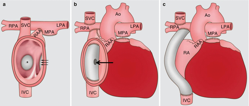
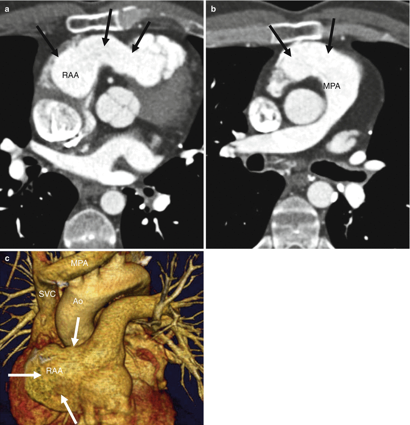
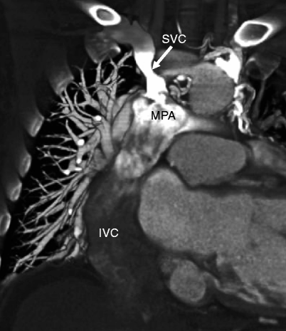
Fig. 25.5
Fontan procedures. Panel (a) demonstrates the classic Fontan procedure. The right atrial appendage (RAA) is connected to the pulmonary artery. The asterisk (*) marks the closed atrial septal defect. The three arrows point to the over sewn tricuspid valve. Panel (b) shows the lateral tunnel procedure. Systemic blood from the inferior vena cava (IVC) is redirected via an intra-atrial tunnel to the pulmonary arteries. The main pulmonary artery is ligated. The superior vena cava (SVC) is connected to the right pulmonary artery as a bidirectional cavopulmonary anastomosis (bidirectional Glenn shunt). Note the presence of fenestration (arrow) in the interatrial tunnel. Panel (c) illustrates an extracardiac conduit procedure. Systemic blood flow from the IVC is redirected via an extracardiac conduit to the pulmonary arteries. Similar to the intra-atrial tunnel, the SVC is connected to the right pulmonary artery as a bidirectional cavopulmonary anastomosis. Currently, the Fontan circulation is achieved by using a bidirectional Glenn shunt and a lateral tunnel or extracardiac conduit Fontan. Ao aorta, MPA main pulmonary artery, LPA left pulmonary artery, RPA right pulmonary artery
Fig. 25.6
Classic Fontan shunt for tricuspid atresia. Panels (a) and (b) are transaxial views. Panel (c) is a 3D image. The classic Fontan (arrows) from the right atrial appendage (RAA) to the main pulmonary artery (MPA) is depicted. In panel (c), note the superior vena cava (SVC) which is surgically connected to the right pulmonary artery (the anastomosis is posterior and thus not visualized in this image)
Fig. 25.7


Lateral tunnel Fontan for tricuspid atresia. A coronal 3D image shows the opacified superior vena cava (SVC) and unopacified inferior vena cava (IVC), both of which are connected to the main pulmonary artery (MPA). The tunnel is created within the right atrium using prosthetic material. RA right atrium
Stay updated, free articles. Join our Telegram channel
Full access? Get Clinical Tree
