SECTION 1
CLINICAL CASE PRESENTATION
A 73-year-old white man with a past medical history of coronary artery disease, status post 4-vessel coronary bypass surgery in 1997, Type 2 diabetes mellitus, hypertension, and diverticular disease presented to our hospital with a chief complaint of “shortness of breath.” For 2 weeks, he had been complaining of dyspnea on exertion (DOE), a nonproductive cough, and chest heaviness that had progressively worsened. In the emergency department, he was hypoxic (requiring oxygen via a non-rebreather mask), tachycardic, and hypotensive with a systolic blood pressure (SBP) in the 80s that did not respond to intravenous (IV) fluids.
On examination, he was obese and appeared quite uncomfortable, only able to speak 4 to 5 words at a time. His cardiac exam showed significant tachycardia, a normal S1 and S2 with a prominent P2 at the left sternal border at the 4th intercostal space and a right parasternal heave. Pulmonary examination revealed that he had decreased breath sounds at the bases bilaterally with poor chest expansion during inspiration. Extremity exam was significant for unilateral left lower leg 1+ pitting edema. Otherwise, the rest of the physical exam was unremarkable.
Laboratory results were significant for brain natriuretic peptide (BNP) 3382, troponin 5.3, and an arterial blood gas (ABG) of pH 7.2, PCO2 30, PO2 104, and HCO3 11 on 100% oxygen. An electrocardiogram (ECG) showed an S wave in lead I, a Q wave in lead III, and an inverted T wave in lead III (Figure 11-1-1). A transthoracic echocardiogram (TTE) showed a large thrombus in the pulmonary arterial trunk that extended into the pulmonary arteries (PA) (Figure 11-1-2). The echocardiogram showed a moderately dilated right ventricle (RV) with severe systolic dysfunction, a flattened septum due to RV pressure overload, and a right ventricular systolic pressure (RVSP) of 51 mm Hg consistent with an acute pulmonary embolism (PE) (Figure 11-1-3). He underwent a computed tomography (CT) scan that confirmed large bilateral pulmonary emboli (PE) (Figure 11-1-4). Figure 11-1-5 demonstrates the thrombi removed at surgery.

FIGURE 11-1-1 Classic ECG findings in acute PE with demonstration of sinus tachycardia, S wave in lead I, Q wave in lead III, and an inverted T wave in leads III and V3.

FIGURE 11-1-2 Two-dimensional image on TTE of a large embolism in the main pulmonary artery in the parasternal short-axis window.

FIGURE 11-1-3 Two-dimensional image on TTE in the parasternal short-axis (upper panel) and subcostal views (lower panel). This demonstrates a dilated, hypokinetic right ventricle (RV) with signs of pressure overload (flattened septum in systole).

FIGURE 11-1-4 Computed tomography imaging of the massive bilateral PEs (arrows). The four images demonstrate the large thrombus in the main and left PA (upper left), in the right PA (upper right), in the main and both PAs (lower left), and in the more distal right PA (lower right).

FIGURE 11-1-5 Demonstration of the pulmonary arterial clots in the operating room postembolectomy (A) and size comparison with a finger that measures 5 cm (B).
CLINICAL FEATURES
• Most patients with a pulmonary embolism (PE) present with dyspnea and/or chest pain. Less commonly, a patient may present with cyanosis, syncope, or hemoptysis.4
• Tachycardia is found in an average of 43% of patients diagnosed with PE.
• Typically, the remainder of the examination is normal, but in patients with massive or submassive PE, signs of RV dysfunction or failure such as hypotension, elevated jugular pressure, a RV S3 gallop, a prominent P2, and a left parasternal heave may be seen and can help narrow the diagnosis.4
EPIDEMIOLOGY
• The overall average incidence of acute PE is between 23 and 69 per 100 000.4,5 Of these, 4.2% are massive PE, causing systemic hypotension or shock.6 The mortality rate from PE is greater than 15% within the first 3 months after diagnosis in patients who are hemodynamically stable and up to 58% in those who present with hemodynamic instablility.6
ETIOLOGY AND PATHOPHYSIOLOGY
• Venous thrombi are typically formed in the deep veins of the calf, often arising in sites of venous stasis. From the deep veins of the calf, the thrombus will propagate to the proximal veins and, from there, embolize, traversing the inferior vena cava (IVC), right atrium (RA), RV, and finally dwelling in the pulmonary arteries (PA). Depending on the size of the embolus, it may cause obstruction in the main PA down to the subsegmental pulmonary arteries.
• Massive PE is defined as an embolism that causes shock, SBPs less than 90 mm Hg, or a drop in SBP greater than 40 mm Hg for greater than 15 minutes.7 A submassive PE is an embolism that causes RV dysfunction without systemic arterial hypotension.
• In patients with either a submassive or massive PE, obstruction of the main PA or its branches along with increased pulmonary vascular resistance (PVR) from hypoxic vasoconstriction leads to increased pulmonary arterial pressure (PAP) and RV afterload.8 The RV wall tension will rise, leading to RV enlargement, dysfunction, and failure. RV enlargement will cause a leftward shift of the interventricular septum and lead to reduced left ventricular (LV) filling. The combination of RV failure and decreased LV preload will ultimately lead to hemodynamic instability.8
• Inherited risk factors for PE include Factor V Leiden, prothrombin gene mutation, and deficiencies of antithrombin III, protein C, or protein S.7 Acquired risk factors include conditions that cause alterations in Virchow’s triad (endothelial injury, hypercoaguability, and venous stasis) such as cancer, congestive heart failure (CHF), chronic obstructive pulmonary disease (COPD), myocardial infarction (MI), obesity, aging smoking, stroke (CVA), oral contraceptive pills or hormone replacement, family history of venous thromboembolism, and recent surgery or trauma.5
ECHOCARDIOGRAPHY
• Patients presenting with symptoms and signs of PE do not typically undergo echocardiography for diagnosis. However, if a patient is hemodynamically unstable and/or is unable to undergo spiral CT, transthoracic echocardiography (TTE) should be performed if a PE is suspected, especially if the patient presents with unexplained dyspnea, syncope, or right ventricular failure.9
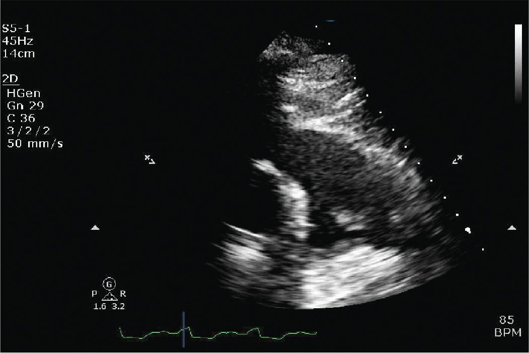
• Echocardiographic assessment, rather than being employed for diagnosis, is utilized to assess the hemodynamic consequences of PE and thus can further assist in guiding therapy. Figures 11-1-6 to 11-1-9 from other patients demonstrate additional echocardiographic images in patients with a PE.
• In addition, following therapy for acute PE, echocardiography is employed to evaluate RV function and PAP and in surveillance of cardiac function and pulmonary pressures following a PE for long-term effects.
• With echocardiography, a thrombus may be visualized in the main PA or in the proximal portions of the right or left PA. The presence of a RA or RV thrombus may also be seen. The sensitivity for detecting PA thrombus is improved with the use of transesophageal echocardiography (TEE).
• In addition to directly visualizing a PE on echocardiography, indirect signs of PE causing acute cor pulmonale may be seen as well. These will be detailed later in this section.
![]() Pulmonary hypertension causing increased RV afterload. PAP is detected by Doppler flow velocity recording of the RV outflow tract. In a normal patient, peak ejection velocity is monophasic and occurs during midsystole. However, in patients with pulmonary hypertension, the ejection profile may be biphasic with a reduction in midsystolic velocity.9
Pulmonary hypertension causing increased RV afterload. PAP is detected by Doppler flow velocity recording of the RV outflow tract. In a normal patient, peak ejection velocity is monophasic and occurs during midsystole. However, in patients with pulmonary hypertension, the ejection profile may be biphasic with a reduction in midsystolic velocity.9
![]() RV enlargement on the apical 4 chamber view. Because the two ventricles are enclosed in the pericardial space, enlargement of the RV will cause a leftward shift of the interventricular septum. The RV end-diastolic to LV end-diastolic ratio will be increased (greater than 0.6). A ratio ranging from 0.6 to 1 indicates mild RV dilatation, whereas a ratio from 1 to 2 indicates severe RV dilatation.9
RV enlargement on the apical 4 chamber view. Because the two ventricles are enclosed in the pericardial space, enlargement of the RV will cause a leftward shift of the interventricular septum. The RV end-diastolic to LV end-diastolic ratio will be increased (greater than 0.6). A ratio ranging from 0.6 to 1 indicates mild RV dilatation, whereas a ratio from 1 to 2 indicates severe RV dilatation.9
![]() Septal flattening and paradoxical septal motion may be seen. Normally, the interventricular septum thickens during systole and moves toward the LV. In a patient with PE causing acute cor pulmonale, RV contraction is prolonged and the interventricular septum moves toward the LV during diastole.9
Septal flattening and paradoxical septal motion may be seen. Normally, the interventricular septum thickens during systole and moves toward the LV. In a patient with PE causing acute cor pulmonale, RV contraction is prolonged and the interventricular septum moves toward the LV during diastole.9
![]() McConnell sign. This occurs when there is appearance of normal wall motion of the apex of the RV accompanied by abnormal wall motion or hypokinesis of the RV free wall.10
McConnell sign. This occurs when there is appearance of normal wall motion of the apex of the RV accompanied by abnormal wall motion or hypokinesis of the RV free wall.10
![]() Diastolic LV impairment. Because the two ventricles are enclosed within the pericardium, the sum of diastolic ventricular dimensions remains constant. Therefore, with acute RV dilatation and failure, LV volume may be reduced and LV filling may be impaired resulting in a further reduction in LV dimension.9
Diastolic LV impairment. Because the two ventricles are enclosed within the pericardium, the sum of diastolic ventricular dimensions remains constant. Therefore, with acute RV dilatation and failure, LV volume may be reduced and LV filling may be impaired resulting in a further reduction in LV dimension.9
![]() RV hypertrophy. In acute cor pulmonale, the RV should not demonstrate hypertrophy. However, in patients with chronic cor pulmonale, RV hypertrophy will be present. Therefore, a lack of RV hypertrophy can help differentiate acute cor pulmonale from chronic pulmonary hypertension.9
RV hypertrophy. In acute cor pulmonale, the RV should not demonstrate hypertrophy. However, in patients with chronic cor pulmonale, RV hypertrophy will be present. Therefore, a lack of RV hypertrophy can help differentiate acute cor pulmonale from chronic pulmonary hypertension.9

FIGURE 11-1-6 Parasternal long axis demonstrating a massively dilated RV that is severely hypokinetic from a patient presenting in shock from a massive PE.

FIGURE 11-1-7 RV inflow view demonstrating a large thrombus in transit extending from the IVC into the RA and across the TV into the RV.

FIGURE 11-1-8 Apical 4 chamber view from the patient in Figure 11-1-7 again demonstrating a large thrombus in the RA and RV. The RV is dilated as well. There is a pericardial effusion present, most likely related to this patient’s lung cancer.

FIGURE 11-1-9
Stay updated, free articles. Join our Telegram channel

Full access? Get Clinical Tree


