Cardiogenic Shock

Chapter 19


Cardiogenic Shock


1. Define cardiogenic shock.


    Cardiogenic shock is a state of end-organ hypoperfusion due to cardiac failure and the inability of the cardiovascular system to provide adequate blood flow to the extremities and vital organs. In general, patients with cardiogenic shock manifest persistent hypotension (systolic blood pressure less than 80 to 90 mm Hg or a mean arterial pressure 30 mm Hg below baseline) with severe reduction in cardiac index (less than 1.8 L/min/m2) in the presence of adequate or elevated filling pressure (left ventricular end-diastolic pressure more than 18 mm Hg or right ventricular end-diastolic pressure more than 10 to 15 mm Hg).


2. What are the various types of shock?


    Blood flow is determined by three entities: blood volume, vascular resistance, and pump function. There are three main types of shock: (1) hypovolemic, (2) vasogenic or distributive, and (3) cardiogenic. Examples of causes of hypovolemic shock include gastrointestinal bleeding, severe hemorrhage, and severe diabetic ketoacidosis (as a result of volume depletion). Examples of vasogenic shock include septic shock, anaphylactic shock, neurogenic shock, and shock from pharmacologic causes. There are many causes of cardiogenic shock, although acute myocardial infarction (MI) is the most common. Cardiogenic shock can be separated into true cardiac causes, such as MI, and extracardiac causes, such as obstruction to inflow (tension pneumothorax, cardiac tamponade) or outflow (pulmonary embolus).


3. Describe the clinical signs observed in cardiogenic shock and other types of shock?


    The medical history and clinical examination help in making the diagnosis of cardiogenic shock. Feeling the extremities and examining the jugular veins provide vital clues: warm skin is suggestive of a vasogenic cause; cool, clammy skin reflects enhanced reflex sympathoadrenal discharge leading to cutaneous vasoconstriction, suggesting hypovolemia or cardiogenic shock. Distended jugular veins, rales, and an S3 gallop suggest a cardiogenic cause rather than hypovolemia. Figure 19-1 presents an algorithm for the evaluation and treatment of cardiogenic shock.
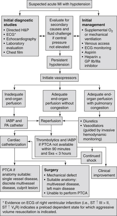


    It is important to note that the clinical examination and chest radiograph may not be reliable predictors of the pulmonary capillary wedge pressure (PCWP). Neither clinically reflect an elevated PCWP in up to 30% of cardiogenic shock patients. In addition, both cardiac tamponade and massive pulmonary embolism can present as cardiogenic shock without associated pulmonary congestion. Right-sided heart catheterization with intracardiac pressure and cardiac pressure measurements is important to confirm the diagnosis of cardiogenic shock.


4. Do all patients with cardiogenic shock have an increased heart rate?


    No. Patients with cardiogenic shock related to third-degree heart block or drug overdose (such as β-blockers and calcium channel antagonists overdose) can present with bradycardia and require temporary transvenous pacemaker implantation.


5. What are the determinants of central venous pressure (CVP)?


    The normal CVP is 5 to 12 cm H2O. Intravascular volume, intrathoracic pressure, right ventricular function, and venous tone all affect the CVP. To reduce variability caused by intrathoracic pressure, CVP should be measured at the end of expiration.


6. What is the significance of a loud holosystolic murmur in a patient with shock after acute myocardial infarction?


    New loud holosystolic murmurs with MI indicate either papillary muscle rupture or an acute ventricular septal defect (VSD). These may be indistinguishable, but acute VSD usually occurs with an anteroseptal MI and has an associated palpable thrill. Papillary rupture often does not have a thrill and is usually seen in inferior MI. These often cause shock on the basis of reduced forward blood flow and can be differentiated by echocardiography or pulmonary artery catheterization. Both require emergent cardiothoracic surgery for early repair. Note that in some patients, particularly those who develop acute mitral regurgitation, the murmur may be soft or inaudible (as a result of a small pressure gradient between the left ventricle and left atrium [or right ventricle]).


7. How can one differentiate cardiogenic from septic shock?


    In classic septic shock, the systemic vascular resistance (SVR) and the PCWP are reduced and the cardiac output is increased. These are usually opposite to the findings in cardiogenic shock. However, a significant decrease in cardiac output may occur in advanced and late stages of sepsis (cold septic shock, which carries a very high mortality rate). Many patients with cardiogenic shock may have normal SVR (i.e., relatively low), even while on vasopressor therapy. Patients with cardiogenic shock may also become dry (normal or low PCWP) with overzealous diuresis. Conversely, septic shock patients may become wet (high PCWP) with overzealous volume replacement. It is therefore ill advised to depend solely on the aforementioned hemodynamic criteria to differentiate cardiogenic shock from septic shock.


8. What is the most common cause of cardiogenic shock?


    Acute MI remains the leading cause of cardiogenic shock in the United States. In fact, despite the decline in its incidence with progressive use of timely primary percutaneous coronary intervention (PCI), cardiogenic shock still occurs in 5% to 8% of hospitalized patients with ST segment elevation myocardial infarction (STEMI). Unlike what is commonly believed, cardiogenic shock may also occur in up to 2% to 3% of patients with non–ST segment elevation myocardial infarction (NSTEMI). Overall, 40,000 to 50,000 cases of cardiogenic shock occur annually in the United States.


9. Describe the pathophysiology of cardiogenic shock among patients with acute myocardial infarction?


    Left ventricular (LV) pump failure is the primary insult in most forms of cardiogenic shock. The degree of myocardial dysfunction that initiates cardiogenic shock is often, but not always, severe. Hypoperfusion causes release of catecholamines, which increase contractility and peripheral blood flow, but this comes at the expense of increased myocardial oxygen demand and its proarrhythmic and cardiotoxic effects. The decrease in cardiac output also triggers the release of vasopressin and angiotensin II, which lead to improvement in coronary and peripheral perfusion at the cost of increased afterload. Neurohormonal activation also promotes salt and water retention, which may improve perfusion but exacerbates pulmonary edema.

Stay updated, free articles. Join our Telegram channel

Jun 5, 2016 | Posted by in CARDIOLOGY | Comments Off on Cardiogenic Shock

Full access? Get Clinical Tree

Get Clinical Tree app for offline access